Can a Frog Peptide Heal Skin Wounds? What the OL-RA11 Autophagy Study Actually Shows
The OL-RA11 frog peptide wound healing study is promising preclinical research, not proof that OL-RA11 heals wounds in people. Researchers identified a new 11-amino-acid peptide from the frog Odorrana livida, tested it in cells, and reported faster repair in a full-thickness skin-wound model in mice. No human participants received OL-RA11.
The paper’s main contribution is a biologically coherent, two-cell explanation. In the inflammatory stage, OL-RA11 was associated with restored autophagy in macrophages, a shift toward M2-associated markers, and lower expression of several pro-inflammatory cytokines. In the proliferative stage, the peptide was associated with Wnt/β-catenin signaling in keratinocytes, greater migration and proliferation, and more re-epithelialization.
Quick answer: OL-RA11 improved several cell, pathway, and wound-model outcomes in one preclinical study. The work strengthens biological plausibility, but it does not establish human efficacy, human safety, an effective way to administer the peptide, durability, or superiority to existing wound care.
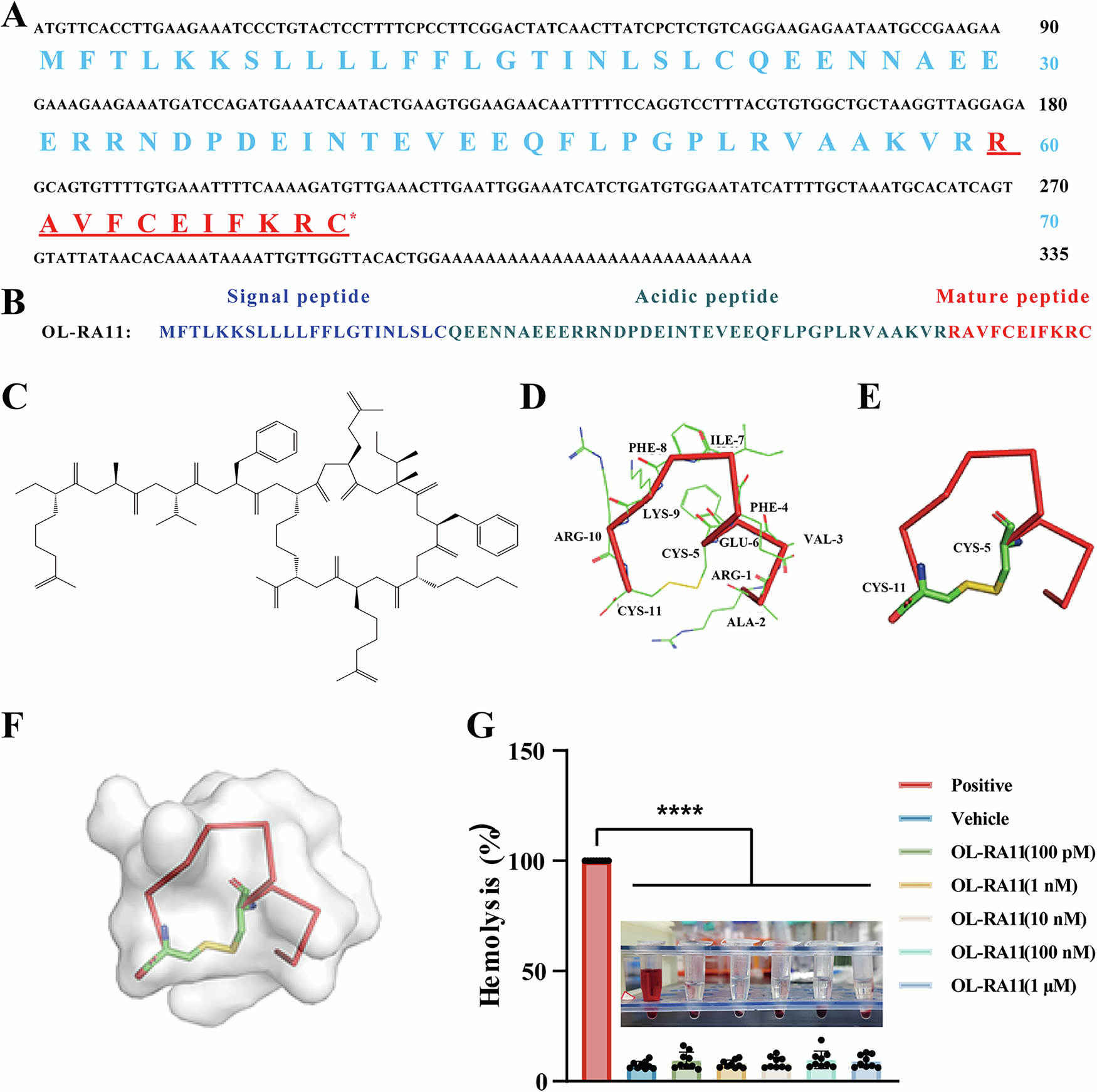
The authors derived the mature OL-RA11 sequence from an Odorrana livida skin cDNA library and predicted a disulfide bond between its two cysteines. Figure adapted without modification from Fu et al., 2026, under CC BY 4.0.
What is OL-RA11?
OL-RA11 is a newly described, frog-derived research peptide—not an approved wound treatment. The researchers screened a previously constructed Odorrana livida skin cDNA library and inferred the mature sequence RAVFCEIFKRC from a 70-amino-acid precursor. They reported no matching previously described sequence in a database search.
The “11” refers to the mature peptide’s 11 amino acids. Structural modeling predicted a disulfide bond between cysteine 5 and cysteine 11. In laboratory stability testing, the cyclic form degraded more slowly than a linear version lacking that bond, particularly at higher temperatures.
That is peptide discovery and characterization. It establishes identity, sequence, a predicted structural feature, and comparative stability under laboratory conditions. It does not establish what the peptide will do in a human body.
What did the OL-RA11 study actually test?
The study combined three evidence layers that should not be blurred together: peptide discovery, cell/pathway experiments, and mouse wound outcomes. Each answers a different question.
| Evidence layer | What the researchers did | What it can support | What it cannot establish | |---|---|---|---| | Peptide discovery | Screened a frog skin cDNA library, inferred the mature sequence, synthesized OL-RA11, and compared cyclic, linear, and scrambled forms | OL-RA11 is a newly reported sequence with structure-dependent laboratory activity | Human wound healing or clinical safety | | Cell and pathway experiments | Used mouse RAW264.7 macrophages, human HaCaT keratinocytes, cytokine assays, migration/proliferation assays, pathway proteins, autophagy reporters, inhibitors, and microscopy | A plausible mechanism involving macrophage autophagy and keratinocyte Wnt signaling | Whether the same effects occur at useful exposures in people | | Wound-model outcomes | Applied OL-RA11 topically in a full-thickness excisional wound model in male Kunming mice | Improved closure and histologic repair in that model | Human efficacy, durability, comparative clinical benefit, or a treatment protocol |
The mouse experiment reported day-7 wound-closure rates of 78.1%, 82.5%, and 83.9% across three OL-RA11 concentrations, versus 61.2% with vehicle. The study also reported more re-epithelialization and changes in granulation tissue, collagen organization, and inflammatory markers. Those are meaningful model outcomes, but they remain outcomes in experimentally wounded mice.
How did OL-RA11 affect macrophages and autophagy?
The macrophage evidence supports an autophagy-linked anti-inflammatory mechanism, although it does not prove a human therapeutic effect. In LPS-stimulated mouse macrophages, OL-RA11 reduced IL-6, IL-8, and TNF-α release. The intact peptide was more active than a linear form, while a scrambled control was inactive in key assays.
The researchers then examined autophagic flux rather than relying on one marker alone. They reported:
- lower phosphorylation of PI3K, AKT, and mTOR;
- higher LC3B and lower p62 levels;
- more autolysosome formation in an mCherry-GFP-LC3B reporter assay;
- ultrastructural evidence consistent with improved autophagosome-lysosome fusion; and
- weaker anti-inflammatory effects when autophagy was blocked with bafilomycin A1 in cells.
In wound tissue, OL-RA11 was also associated with more LC3 signal in F4/80-positive macrophages, higher ARG1 and IL-10, and lower iNOS, IL-6, IL-1β, and TNF-α. The authors interpreted this pattern as increased autophagy supporting M2 polarization and inflammation resolution.
“M2” is useful shorthand here, but macrophage states in living tissue are more complex than a clean M1-versus-M2 switch. Marker changes support the proposed direction; they are not a complete map of macrophage behavior.
Mechanism versus outcome: Blocking autophagy weakened several OL-RA11 effects, which makes autophagy more than a coincidental marker. It still does not show that manipulating this pathway with OL-RA11 will safely improve human wounds.
What was the second stage involving keratinocytes and Wnt signaling?
The second proposed stage is regenerative rather than primarily anti-inflammatory. Keratinocytes help rebuild the epidermal barrier by proliferating and moving across the wound surface. In HaCaT keratinocyte experiments, OL-RA11 increased proliferation and migration, while scrambled and linear controls were less active or inactive.
RNA sequencing of mouse wound tissue pointed to several pathways, including Wnt signaling. Protein experiments then showed higher nuclear β-catenin and higher expression of downstream proteins Cyclin D1 and c-Myc in treated wound tissue and keratinocytes. The authors linked those changes to migration, proliferation, and re-epithelialization.
This is a reasonable pathway chain: OL-RA11 exposure, Wnt/β-catenin-associated changes, keratinocyte behavior, then epidermal repair in the model. But the study did not identify a human dose-response relationship, a validated molecular target for OL-RA11, or clinical benefit.
Wnt/β-catenin signaling also appears in a COPD study of elastin-derived peptides and alveolar repair, but there it was linked to impaired repair rather than improved skin healing, underscoring why tissue context and pathway direction matter.
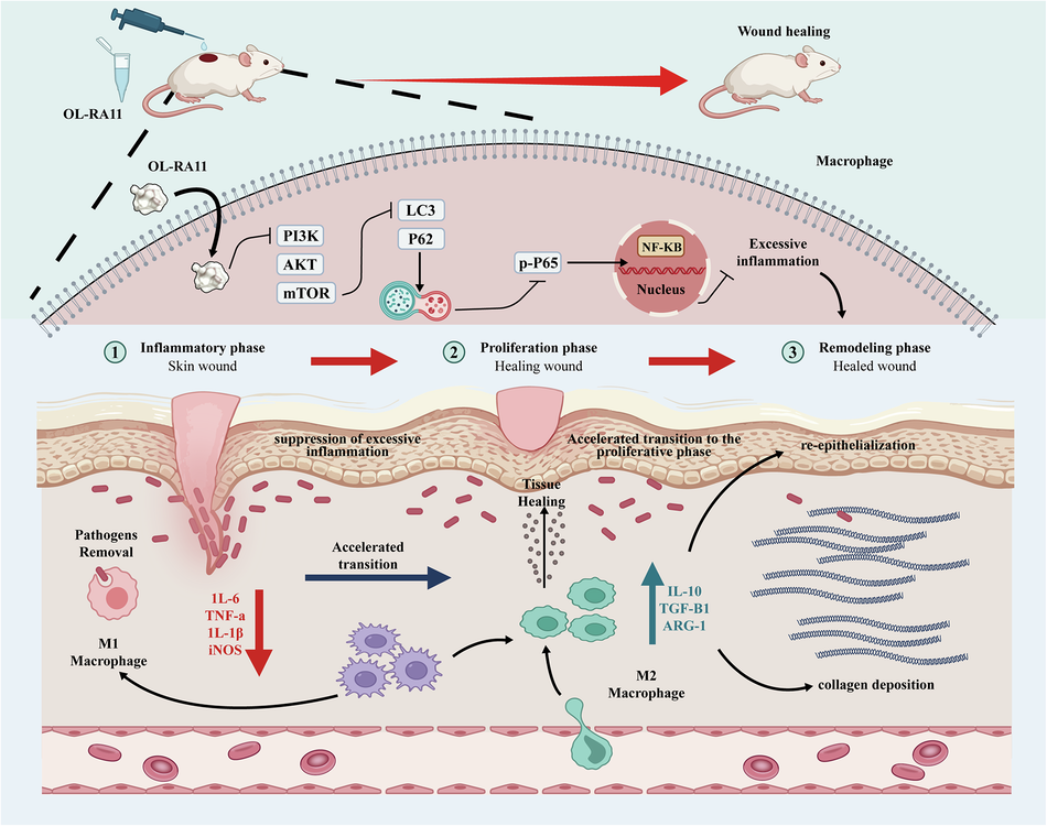
The authors propose that OL-RA11 coordinates inflammation resolution with later tissue repair. This is a mechanistic model built from preclinical experiments, not a clinical pathway validated in patients. Figure adapted without modification from Fu et al., 2026, under CC BY 4.0.
Why does a two-cell mechanism strengthen plausibility without proving efficacy?
A coherent dual-cell mechanism is stronger than a single unexplained outcome because multiple observations point in the same biological direction. The macrophage work addresses how excessive inflammation might resolve; the keratinocyte work addresses how the surface barrier might be rebuilt. Inhibitor experiments, pathway measurements, cell behavior, and wound histology provide partial cross-checks.
The comparison with rapamycin is also conceptually useful. Autophagy induction alone improved some repair measures but did not reproduce all of the reported tissue-quality effects of OL-RA11. The authors argue that resolving inflammation is not enough; repair also needs a proliferative signal.
Still, coherence can become seductive. A mechanism may be internally consistent and still fail clinically because:
- the relevant concentration cannot be maintained safely in human tissue;
- metabolism, delivery, or tissue penetration differs between mice and people;
- the pathway effect is not specific enough;
- short-term closure does not translate into durable, functional repair;
- results change in infected, diabetic, ischemic, chronic, or otherwise complex wounds; or
- benefits do not exceed those of established care.
Biological plausibility raises confidence that an observation is worth pursuing. Clinical efficacy asks a separate question and requires human trials.
What are the biggest translation limits?
The largest limitation is simple: this was not a human wound-healing trial. The full experimental report and supplements leave several important questions unanswered.
Human safety is unknown
The study reported no hemolysis in mouse red blood cells and no mortality, major-organ histopathology, or abnormal weight trajectory during short mouse toxicity experiments. Those findings are useful for early screening. They cannot establish local irritation, immunogenicity, infection risk, systemic exposure, reproductive risk, long-term toxicity, or safety in people with medical conditions.
Administration is unresolved
The wound model used topical experimental application under controlled conditions. That does not establish a human formulation, delivery system, effective exposure, dosing schedule, or stability in a real wound environment. This article does not turn animal methods into self-treatment instructions.
The model was narrow
The in-vivo wound experiments used male mice. The authors explicitly noted that this prevented assessment of sex differences and called for studies in both sexes and larger animals such as pigs. Experimentally created acute wounds also differ from many chronic human wounds.
Durability and comparative benefit remain unknown
Day-7 closure and histology do not establish durable barrier function, scar quality, recurrence, infection outcomes, or long-term performance. The study included laboratory comparators, but it was not designed to compare OL-RA11 against modern human wound-care standards in a clinical setting.
Target specificity remains uncertain
The authors acknowledged that in-vivo stability, biological activity, and target specificity have not been characterized. Effects on fibroblasts, endothelial cells, and communication between macrophage autophagy and keratinocyte Wnt signaling also remain open questions.
Does this study say anything about BPC-157, TB-500, or cosmetic peptides?
No. OL-RA11 is a distinct sequence studied in a distinct experimental program. The paper does not test BPC-157, TB-500, GHK-Cu, cosmetic peptides, supplements, or “research-use-only” products. Shared marketing words such as “repair” or “healing” do not create scientific equivalence.
For context, see What Is BPC-157?, What Is TB-500?, and What Is GHK-Cu?. Our guide to what “preclinical” actually means explains why animal and cell findings cannot be treated as human treatment evidence. Research-Use-Only Peptides: What the Label Means covers a separate regulatory and evidence issue.
What evidence would move OL-RA11 closer to clinical relevance?
The next steps are replication, better translational models, pharmacology, and eventually carefully staged human research—not consumer experimentation. Useful evidence would include:
- independent replication of the cell and mouse findings;
- target-identification and cell-specific genetic experiments;
- pharmacokinetic, tissue-penetration, stability, and immunogenicity studies;
- both-sex studies and models of chronic, infected, diabetic, or ischemic wounds;
- larger-animal wound models with functional and longer-term outcomes;
- formulation and manufacturing work; and
- phased human trials designed first around safety and exposure, then efficacy and comparative benefit.
Until those steps exist, OL-RA11 is best understood as an interesting research lead.
Frequently asked questions
Did OL-RA11 heal wounds in humans?
No. The study used cells and a full-thickness wound model in mice. It reported no human trial.
Was OL-RA11 discovered directly in frog skin secretions?
The researchers identified the sequence by screening a previously constructed Odorrana livida skin cDNA library, then chemically synthesized the mature peptide for experiments.
Does the study prove autophagy caused every wound benefit?
No. Inhibitor and flux experiments strengthen the case that autophagy contributed to the macrophage and wound effects. The study also proposed a separate Wnt-linked keratinocyte pathway, and several upstream targets and cell interactions remain unresolved.
Is OL-RA11 safe for topical or injected human use?
That has not been established. Short mouse and laboratory safety observations cannot determine human safety, and this article provides no administration or self-treatment guidance.
Is OL-RA11 related to BPC-157 or TB-500?
The study establishes no such relationship. They are different peptides with different sequences and evidence bases.
Sources
- Fu Z, Xiao Z, Li Y, et al. “A frog bioactive peptide suppresses inflammation by modulating autophagy for skin wound regeneration.” Communications Biology. Published August 12, 2026. doi:10.1038/s42003-026-10766-z
- Fu Z, Xiao Z, Li Y, et al. Supplementary Information for the same article. Publisher supplement
This article is educational. It does not diagnose wounds or provide treatment, dosing, injection, topical-use, sourcing, or purchasing instructions.