TR-MOD: What a Biofilm-and-Fibroblast Lab Study Can—and Cannot—Show About a “Wound-Healing” Peptide
The short answer: The October 7, 2026 TR-MOD peptide wound healing study is an in-vitro proof of concept, not evidence that TR-MOD heals human wounds, treats an infection, eradicates a mature biofilm, or is safe in people. The researchers found antibacterial activity against four Pseudomonas aeruginosa strains, less crystal-violet-stained material when the peptide was present during biofilm formation, and faster gap closure by cultured human dermal fibroblasts. Those are distinct laboratory signals. None is a clinical outcome.
The open-access Scientific Reports paper by Park and colleagues is scientifically interesting because it asks whether one designed peptide can affect both a pathogen and a host-cell process. It is also a useful lesson in evidence boundaries. A minimum inhibitory concentration is not infection clearance. Reduced biofilm biomass is not proof that embedded bacteria were eradicated. A fibroblast migration assay is not a wound closing in an animal or patient.
For the wider evidence hierarchy, see what “preclinical” actually means.
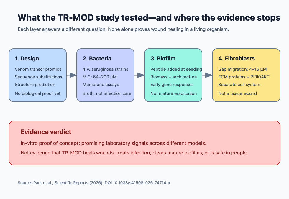
The paper links several laboratory layers, but the clinical bridge remains untested. It did not include a mature-biofilm eradication experiment, an angiogenesis assay, an animal wound model, or a human trial.
Quick verdict: what did the TR-MOD study actually show?
It showed a collection of preclinical signals under controlled laboratory conditions.
| Experimental layer | What was tested | What the result supports | What it does not establish | |---|---|---|---| | Computational design | Sequence, charge, hydrophobicity, amphipathicity, structure and activity predictions | A rationale for selecting TR-MOD | Real-world activity, selectivity or safety | | Planktonic bacteria | MIC and membrane assays in four P. aeruginosa strains | Growth inhibition and membrane effects in broth | Treatment of an infected wound | | Biofilm formation | Peptide added when bacteria were seeded; biomass and architecture assessed later | Interference with biofilm development | Eradication of a mature, established biofilm | | Fibroblast gap assay | HDFα cells at 4, 8 and 16 µM for up to 24 hours | Increased migration-associated gap closure | Tissue repair, re-epithelialization or wound closure in vivo | | Protein/signaling assays | ECM and migration-associated proteins; PI3K/AKT and inhibitor experiments | A pathway hypothesis in cultured fibroblasts | Angiogenesis or a complete wound-healing mechanism | | Cytocompatibility | Three human cell types and bovine red blood cells | Short-term tolerability within tested assay conditions | Human local or systemic safety |
The paper was published as an early, citable accepted version. Its own discussion calls for in-vivo infection and wound-healing models before therapeutic conclusions.
What is TR-MOD, and how was it designed?
TR-MOD is a 16-amino-acid experimental antimicrobial peptide designed from a spider venom-gland transcriptomic lead. The researchers compared whole-body and venom-gland RNA-sequencing data from Araneus ventricosus, selected 25 sequences enriched more than five-fold in venom gland tissue, and ran 19 eligible sequences through antimicrobial-peptide prediction. Two were classified as AMP-like.
Both candidates contained the same 16-residue helical region, named TR-ORI. The team then substituted tryptophan and arginine residues to increase hydrophobicity, positive charge and separation of hydrophobic and charged faces. The resulting sequence, TR-MOD, had a predicted net charge of +6.9 versus +2.9 for TR-ORI and stronger in-silico antimicrobial scores.
The synthesized peptide was reported at 95.09% purity with a measured molecular weight of 2,222.68 Da. Computational tools predicted an amphipathic alpha helix and low toxicity, but those predictions were only the first filter. Prediction tools are useful for prioritizing candidates; they do not validate biological performance.
The transcriptomic discovery step also had a limitation acknowledged by the authors: the number of biological replicates, particularly for venom-gland samples, was limited.
What did the planktonic antibacterial tests find?
TR-MOD inhibited growth in broth, but the required concentrations were relatively high. The study used one reference strain, P. aeruginosa ATCC 15692, and three multidrug-resistant strains: CCARM 2007, CCARM 2095 and CCARM 2241. Each MIC experiment started with 4 × 10^5 CFU/mL, incubated for 16 hours, and was repeated in three independent experiments.
| Strain | TR-MOD MIC | Approximate mass concentration reported | |---|---:|---:| | ATCC 15692 | 200 µM | 444.54 µg/mL | | MRPA CCARM 2007 | 128 µM | 284.5 µg/mL | | MRPA CCARM 2095 | 64 µM | 142.25 µg/mL | | MRPA CCARM 2241 | 64 µM | 142.25 µg/mL |
The paper compared those values with ampicillin, streptomycin, tetracycline and ciprofloxacin. TR-MOD’s MIC was lower than the tested antibiotics in CCARM 2007 and CCARM 2095, but several antibiotic MICs were not reached within the tested range. That makes the comparison informative within this assay, not a claim that the peptide is clinically superior to antibiotics.
In two representative strains—ATCC 15692 and CCARM 2095—TR-MOD also increased DiSC3(5) and NPN fluorescence at one-half and one times the MIC. Those assays support cytoplasmic-membrane depolarization and increased outer-membrane permeability. They do not identify every molecular target or show how the peptide would behave in wound fluid, tissue or blood.
Did TR-MOD disrupt an established biofilm?
No mature-biofilm eradication experiment was reported. The biofilm experiments added TR-MOD immediately when 2 × 10^5 bacterial cells per well were seeded, then measured material after 18 or 24 hours. That design tests interference with biofilm formation and early architecture, not removal of a biofilm that was allowed to mature before treatment.
The main quantitative assay used crystal violet, which measures retained biofilm-associated biomass. TR-MOD was tested at one-quarter, one-half and one times each strain’s MIC:
- ATCC 15692: 50, 100 and 200 µM;
- CCARM 2007: 32, 64 and 128 µM;
- CCARM 2095: 16, 32 and 64 µM; and
- CCARM 2241: 16, 32 and 64 µM.
The comparison groups included untreated control, 3.5% ethanol, tetracycline and ciprofloxacin. Antibiotic concentrations were strain-specific: ciprofloxacin was 0.25 µg/mL for ATCC 15692 and 2.56 µg/mL for the three resistant strains; tetracycline was 16, 512, 512 and 64 µg/mL, respectively. Where an antibiotic MIC was not reached, the authors used the highest tested concentration.
TR-MOD reduced crystal-violet staining at sub-MIC concentrations across all four strains. The quantitative panels used three independent biological replicates, each with six technical replicates, and reported means ± SEM with exact P values on the graphs.
That is a biofilm-development signal. Crystal violet does not distinguish living bacteria from dead bacteria or matrix material. The study did not report post-treatment viable counts from a mature biofilm, a minimum biofilm eradication concentration, or relapse after peptide removal. Calling this “biofilm eradication” would outrun the experiment.
This distinction is similar to the broader problem covered in how to evaluate peptide claims online: the endpoint has to match the headline.
What did the microscopy and biofilm-gene experiments add?
They added mechanism clues, not a second proof of eradication. Field-emission scanning electron microscopy was performed on ATCC 15692, CCARM 2007 and CCARM 2095 after 24 hours with TR-MOD at one times the MIC. The images showed strain-dependent changes in cell morphology, extracellular polymeric substance (EPS) filaments and biofilm structure.
Confocal staining in ATCC 15692 and CCARM 2095 used ConA, SYPRO Ruby and TOTO-1 to visualize polysaccharides, proteins and extracellular DNA. TR-MOD produced punctate redistribution and, in CCARM 2095, lower overall EPS-associated signals. Quantification used three independent biological replicates.
For transcriptional analysis, ATCC 15692 and CCARM 2095 were exposed during early biofilm formation, and RNA was collected four hours after treatment. RT-qPCR examined sadC, siaD, pelA, pslA, algD and cdrA, normalized to 16S rRNA. TR-MOD reduced sadC in both strains, while several other genes moved in opposite directions between strains. The assay used three biological replicates with technical triplicates.
The consistent sadC decrease is a lead because SadC participates in cyclic-di-GMP synthesis. But the investigators did not directly measure intracellular cyclic-di-GMP or SadC activity. Gene-expression changes are associations with a pathway, not proof that the pathway caused the biomass result.
What did the fibroblast “wound-healing” assay measure?
It measured cultured-cell migration into a manufactured gap. Primary human dermal fibroblasts (HDFα) were grown around a removable two-well insert. After the insert was removed, cells were exposed to 4, 8 or 16 µM TR-MOD and photographed at 0, 12 and 24 hours. ImageJ was used to calculate the percentage of the cell-free area that closed.
Gap closure increased with concentration. The experiment used three independent biological replicates and reported means ± SEM. Separate crystal-violet colony and cell-counting assays found no significant increase in proliferation at the tested concentrations, supporting migration rather than simple cell-number expansion as the main contributor.
This is often called a scratch or wound-closure assay, but no animal or person was wounded. There was no skin barrier, immune response, blood flow, bacterial infection, extracellular matrix complexity or mechanical stress. The clean interpretation is fibroblast migration in vitro, not “TR-MOD healed a wound.”
The assay also did not combine fibroblasts with a live infected biofilm. The bacterial and host-cell findings came from separate experimental systems.
Did the study test angiogenesis or prove a wound-healing pathway?
No angiogenesis assay was reported, and the pathway work remained partial. There were no endothelial-cell tube-formation experiments, blood-vessel measurements, VEGF-focused functional assays, animal histology or perfusion endpoints.
The researchers instead measured proteins associated with extracellular-matrix remodeling and fibroblast activation. At 12 hours, fibronectin increased at 4, 8 and 16 µM, while MMP-9 showed a trend rather than a consistent significant increase. At 24 hours, fibronectin remained elevated at 4 and 8 µM; alpha-SMA, vimentin and N-cadherin also increased in concentration-dependent patterns. Western-blot experiments used three biological replicates.
After four hours, AKT phosphorylation was about 1.5-fold the control level and PIK3CA expression increased. The peptide did not significantly change the tested TGF-beta/SMAD3 or Wnt/beta-catenin readouts. Pharmacological experiments used 16 µM TR-MOD with pictilisib, lenvatinib or erdafitinib. PI3K inhibition suppressed the migration signal, while broader RTK and FGFR inhibition attenuated it and allowed partial recovery with TR-MOD.
Those results support a PI3K/AKT-dependent migration hypothesis with an FGFR-related modulatory role. They do not identify a direct receptor, demonstrate peptide-receptor binding or exclude other pathways. The authors specifically called for receptor knockdown, binding studies, broader RTK profiling and time-resolved phosphorylation work.
What did the cytotoxicity and hemolysis tests show?
TR-MOD appeared compatible with selected cells over 24 hours within limited concentration ranges, but the safety margin was not uniformly wide. A CCK-8 viability assay tested primary HDFα fibroblasts, HaCaT keratinocytes and BEAS-2B bronchial epithelial cells from 1 to 200 µM. Each condition used three biological replicates with ten technical replicates.
The paper reported no significant cytotoxicity up to:
- 200 µM in HDFα fibroblasts;
- 32 µM in HaCaT keratinocytes; and
- 64 µM in BEAS-2B cells.
At higher concentrations, the HaCaT and especially BEAS-2B viability curves declined. “No significant cytotoxicity up to” therefore does not mean universally non-toxic.
Hemolysis was tested with bovine—not human—red blood cells from 1 to 200 µM for one hour. PBS and 0.1% Triton X-100 were the negative and positive controls. Three independent biological replicates found no significant hemolysis up to 200 µM. Because 50% hemolysis was not reached, the authors estimated minimum selectivity indices of greater than 1.0, 1.56, 3.13 and 3.13 across the four bacterial strains. They noted that the margin was relatively limited for ATCC 15692.
A supplementary SDS-PAGE experiment found no obvious loss of the TR-MOD band after incubation in 10% human serum for up to 24 hours. That is a coarse stability readout. It does not establish intact bioactivity, metabolite identity, pharmacokinetics or stability in an actual wound environment.
How strong were the replication and statistics?
The study used repeated experiments, but most quantitative claims rested on three biological replicates. The paper states that experiments were performed in at least triplicate, with assay-specific counts given in methods or figure legends. Data were expressed as means ± SEM and analyzed with one-way ANOVA followed by Tukey’s post-hoc test; P < 0.05 was the significance threshold.
Examples include:
- MIC: three independent experiments;
- biofilm crystal violet: three biological × six technical replicates;
- biofilm-gene RT-qPCR: three biological × three technical replicates;
- cell viability: three biological × ten technical replicates;
- fibroblast migration, hemolysis, EPS imaging and western blots: three biological replicates; and
- membrane assays: three biological × four technical replicates.
Technical replicates improve measurement precision but do not replace independent biological replication. The paper did not report preregistration, blinded image analysis or independent laboratory replication. With many endpoints and a small biological n, exact P values do not remove the need for replication and effect-size scrutiny.
Why is a multifunctional peptide design still interesting?
The design is interesting because infected wounds involve both microbial persistence and damaged host tissue. A molecule that genuinely combines antimicrobial, antibiofilm and host-repair activity could simplify a multi-problem intervention. TR-MOD also generated internally connected hypotheses: membrane perturbation, altered biofilm development, fibroblast migration and PI3K/AKT signaling.
But multifunctionality can also multiply failure points:
- Potency: MICs of 64–200 µM are high enough that further optimization is needed, as the authors acknowledge.
- Stability: one gel-based serum experiment cannot predict proteolysis, loss of activity or shelf behavior.
- Delivery: an active concentration in broth or culture medium may not be achievable throughout an infected wound or device biofilm.
- Selectivity: microbial membrane activity must be separated from damage to keratinocytes, airway cells, blood cells and other tissues.
- Resistance and adaptation: the paper did not test serial exposure, resistance emergence or combination durability.
- Manufacturing: a 95.09%-pure research batch is not a validated clinical manufacturing process.
- Model validity: planktonic broth, developing biofilms and fibroblast monolayers do not reproduce an infected wound.
- Mechanism: c-di-GMP and receptor claims remain indirect and partly strain-dependent.
- Translation: there was no animal efficacy, tissue distribution, immunogenicity, toxicology or human evidence.
For another example of why promising antimicrobial-peptide data can remain far from clinical proof, see the 2026 antimicrobial cyclic peptide study.
Bottom line: does TR-MOD heal wounds?
No available experiment shows that TR-MOD heals wounds in animals or people. The paper supports narrower statements: TR-MOD inhibited planktonic P. aeruginosa in broth, interfered with biofilm formation when present from the start, altered selected biofilm and signaling readouts, and increased migration-associated gap closure in cultured dermal fibroblasts.
That makes TR-MOD a preclinical research candidate. The next meaningful evidence would require mature-biofilm challenge models, infected three-dimensional tissue or animal wounds, validated delivery, pharmacology, toxicology, resistance testing and eventually controlled human studies. Until those layers exist, “wound-healing peptide” is a research hypothesis, not a treatment conclusion.
Frequently asked questions
Was TR-MOD tested in humans?
No. The paper contained computational analyses and in-vitro bacterial and human-cell experiments. It included no patient treatment or human safety study.
Did TR-MOD eradicate a mature biofilm?
No. The peptide was added when bacteria were seeded, so the experiments assessed biofilm formation and early architecture. The study did not report viable-count eradication of a pre-established mature biofilm.
Was the fibroblast assay a real wound?
No. It was a removable-insert gap assay in a monolayer of cultured human dermal fibroblasts. Faster cell migration into that gap is not the same endpoint as wound closure in an animal or patient.
Did the study show angiogenesis?
No. It measured fibroblast-associated proteins and signaling but did not report endothelial tube formation, blood-vessel growth, perfusion or other functional angiogenesis endpoints.
Does no cytotoxicity up to 200 µM mean TR-MOD is safe?
No. That threshold applied to HDFα fibroblasts during one 24-hour assay. HaCaT and BEAS-2B cells had lower no-significant-cytotoxicity ranges, and bovine hemolysis does not establish human tissue or systemic safety.
Primary source
- Park HR, Oh JW, Jeong S, et al. Designing a multifunctional peptide with biofilm-disrupting and fibroblast migration-promoting activities for infection management. Scientific Reports. Published October 7, 2026. The full accepted-manuscript PDF and both official supplementary files were reviewed for this article.
This article is for education and evidence interpretation only. It does not provide medical advice, diagnosis, treatment instructions, dosing, formulation guidance, synthesis guidance, purchasing guidance, sourcing advice or self-experimentation guidance.