Can Catestatin Treat Neurodegeneration? What the New Mouse Study Actually Shows
A 2026 study linked catestatin to lower tau, amyloid, and inflammatory readouts in cultures and mouse models—but it did not establish a human treatment.
Can Catestatin Treat Neurodegeneration? What the New Mouse Study Actually Shows
No—catestatin has not been shown to treat neurodegeneration in people. A September 2026 study reported encouraging results across postmortem human brain tissue, neuronal and brain-slice cultures, and two transgenic mouse models. The therapeutic experiments were preclinical, however. They do not establish human efficacy, safety, or brain exposure, nor optimal dosing, durability, or disease modification in people.
The study is still scientifically interesting. Catestatin, or CST, affected several readouts linked to tau, amyloid, inflammation, and behavior. The useful way to read it is as an evidence ladder—not as a treatment announcement.
Quick answer: The paper supports catestatin as a preclinical research candidate for neurodegeneration. It does not support using catestatin as an Alzheimer’s, corticobasal degeneration, progressive supranuclear palsy, or other neurodegenerative-disease treatment.
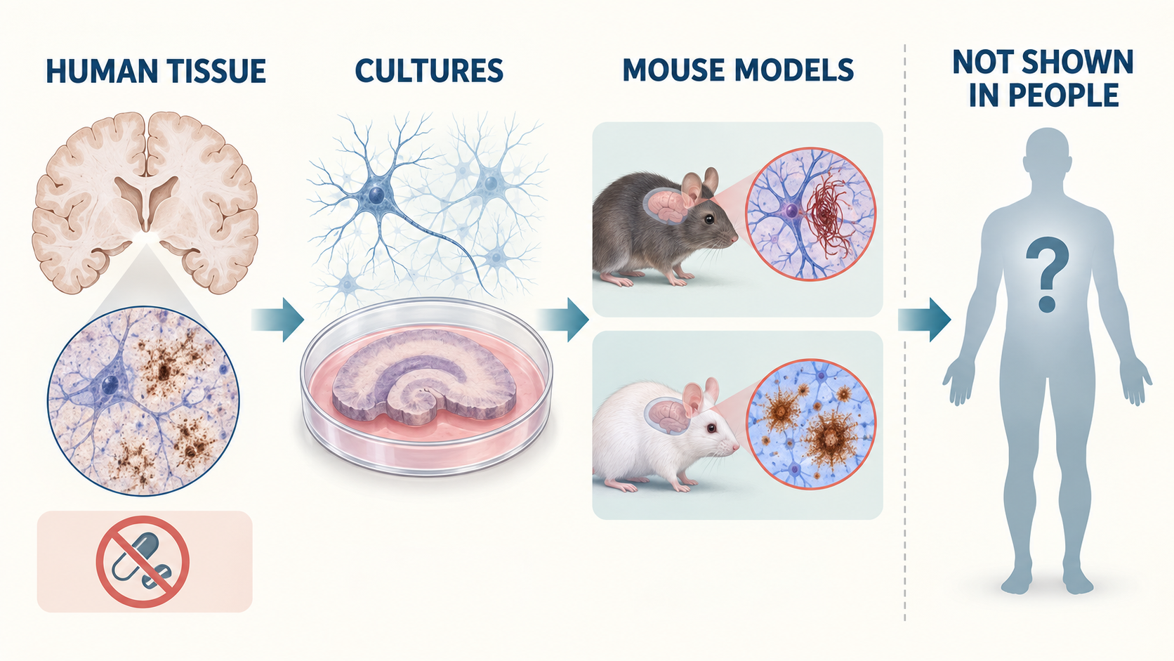
The paper spans four evidence settings, but only the first involved human tissue—and that was observational postmortem analysis, not treatment. The two mouse panels represent distinct tau and amyloid models.
What is catestatin?
Catestatin is a naturally occurring peptide fragment produced from chromogranin A, a protein involved in neuroendocrine secretion. It is also called CST and corresponds to a short region of human chromogranin A.
Catestatin has previously been studied for its ability to inhibit catecholamine release and for possible roles in cardiovascular, metabolic, and immune regulation. That background led the researchers to ask whether altered processing of chromogranin A might also matter in neurodegenerative disease.
The same precursor protein produces other peptides, including pancreastatin. In this study, catestatin and pancreastatin often moved in opposite directions in sampled disease-affected brain regions. That observation is a clue about altered peptide balance. It is not proof that low catestatin causes disease or that replacing it reverses human disease.
What did the catestatin neurodegeneration study find?
The study found a consistent preclinical pattern: lower catestatin in selected human disease samples, less tau pathology in cultured systems given CST, and improved pathological or functional readouts in CST-treated mouse models. Each layer answers a different question.
1. Sampled human brain regions showed an association
The researchers measured CST in postmortem tissue from people with Alzheimer’s disease, corticobasal degeneration, or progressive supranuclear palsy. They reported:
- lower CST in the prefrontal cortex and hippocampal/entorhinal region in advanced Alzheimer’s samples;
- lower CST in frontal cortex samples from corticobasal degeneration;
- lower CST in basal ganglia samples from progressive supranuclear palsy; and
- higher pancreastatin in the sampled Alzheimer’s and corticobasal degeneration regions.
In the Alzheimer’s samples, lower CST also correlated with lower Mini-Mental State Examination scores. Correlation in postmortem tissue cannot show which change came first, rule out disease severity or other confounders, or demonstrate that CST replacement would help living patients.
Evidence boundary: Human tissue can show that a biological feature travels with disease. It cannot, by itself, show that changing that feature treats the disease.
2. Cortical neurons and brain-slice cultures showed lower tau readouts
The researchers then moved from observation to intervention in laboratory models. In cortical neuronal cultures expressing disease-associated P301S tau, CST supplementation reduced measured tau phosphorylation and misfolded tau accumulation. Similar reductions were reported in organotypic hippocampal slice cultures, which preserve more local tissue organization than isolated cells but still remain an ex-vivo model.
These experiments make the biological hypothesis more plausible. They do not reproduce the full physiology, exposure, immune environment, aging process, or clinical complexity of a person with neurodegenerative disease.
3. PS19 mice showed changes in tau pathology, gliosis, and behavior
PS19 mice express mutant human tau and are used to model aspects of tauopathy. In these mice, administered CST was associated with:
- lower pathological tau phosphorylation, misfolding, and seeding-related readouts;
- less microglial and astrocytic activation;
- less hippocampal atrophy in the measured tissues; and
- better performance on selected Y-maze, grip-strength, rotarod, and nesting measures.
Those behavioral tests are legitimately functional outcomes in mice. Calling them proof of restored human cognition would be a category error. A mouse maze result is not a clinical memory outcome, and PS19 biology is not the same as sporadic Alzheimer’s disease, corticobasal degeneration, or progressive supranuclear palsy in people.
4. 5xFAD mice showed lower amyloid and inflammatory readouts
The researchers separately tested 5xFAD mice, a model engineered to develop aggressive amyloid pathology. CST-treated 5xFAD mice had less measured amyloid plaque burden in the hippocampus and cortex and lower markers of microglial and astrocytic activation.
This was not the same experiment as the PS19 study. PS19 is primarily a tauopathy model; 5xFAD is primarily an amyloid model. Using both broadens the preclinical signal, but it does not turn either model into human efficacy evidence.
The distinction matters because a compound can reduce plaques in a transgenic mouse without improving human symptoms or changing the course of Alzheimer’s disease. For a broader guide to this translation gap, see what preclinical actually means.
How might catestatin affect tau and amyloid biology?
The paper proposes that CST restrains an adrenergic stress pathway involving epinephrine and protein kinase A, but this mechanism is not settled human disease biology.
The model is roughly:
- elevated epinephrine increases adrenergic signaling;
- adrenergic signaling activates cyclic-AMP-dependent protein kinase A, or PKA;
- excessive PKA activity can contribute to tau phosphorylation; and
- CST, known to inhibit catecholamine release, may reduce epinephrine and dampen this signaling cascade.
The researchers reported elevated cortical epinephrine in both PS19 and 5xFAD mice, with lower levels after CST treatment. In PS19 tissue and organotypic slice cultures, CST was also associated with less PKA-substrate phosphorylation and less pathological tau phosphorylation. In a slice-culture challenge experiment, CST blunted changes induced by added epinephrine.
That is a coherent mechanistic chain within the tested systems. It does not prove that CST deficiency drives human neurodegeneration, that adrenergic signaling is the only relevant pathway, or that modifying this pathway would produce a net clinical benefit. Catestatin has effects outside the brain, and a mechanism that looks helpful on selected laboratory readouts can still create exposure, off-target, cardiovascular, metabolic, immune, or long-term safety problems.
Mechanism takeaway: Lower epinephrine and reduced PKA hyperactivation are the study’s proposed explanation for part of the CST effect. They are supported by mouse and ex-vivo measurements, not by a human intervention experiment.
Did the study show that catestatin reaches the brain?
It showed brain exposure in a mouse distribution experiment, not in humans. The researchers reported detecting CST in mouse brain tissue after administration and calculated a mouse brain-to-plasma exposure ratio.
That finding is more informative than simply assuming that a peptide crosses into the brain. It still leaves the clinically important questions unanswered: whether a comparable molecule reaches relevant human brain regions, what concentrations are required, how long exposure lasts, whether repeated exposure is safe, and whether the measured tissue signal represents useful target engagement.
Mouse brain distribution should therefore be described as a preclinical pharmacokinetic result—not evidence of human brain delivery.
What does the study not establish?
Nearly every clinical question remains open. The paper does not establish:
- efficacy in people with Alzheimer’s disease, corticobasal degeneration, progressive supranuclear palsy, or another neurodegenerative condition;
- safety or tolerability in humans;
- human pharmacokinetics or brain exposure;
- an effective or optimal human dose, schedule, formulation, or route;
- durability after treatment stops;
- prevention of symptoms or slowing of clinical progression;
- disease modification rather than temporary movement in biomarkers;
- comparative benefit against existing or investigational therapies;
- which patients, if any, might benefit; or
- whether long-term adrenergic, cardiovascular, metabolic, or immune effects would be acceptable.
The study also does not show that low CST is a diagnostic biomarker. Measurements from selected postmortem regions cannot be converted into a clinical test without separate validation.
For help distinguishing a model result from a clinical claim, see peptide research status explained and how to evaluate peptide claims online.
Why the two mouse models should not be blended together
PS19 and 5xFAD supplied complementary but non-interchangeable evidence. The PS19 experiments addressed mutant-tau pathology, gliosis, hippocampal structure, and selected behavioral outcomes. The 5xFAD experiments addressed amyloid plaque burden and neuroinflammation.
The paper did not show, in one model, that CST simultaneously corrected the full human Alzheimer’s disease process. Nor did it establish that a result in a familial amyloid-overexpression model predicts benefit in sporadic Alzheimer’s disease.
This is the same discipline needed when reading other peptide-neurodegeneration studies. For example, the TREM2 cyclic-peptide study stopped at binding and cultured-cell models. The CST paper goes further into animals, but neither study supplies human therapeutic evidence. “Further along the ladder” is not the same as “clinically proven.”
What commercial interests did the authors disclose?
The commercial and intellectual-property context is relevant and should be visible, but it does not invalidate the findings by itself.
The paper reports that senior author Sushil K. Mahata founded CgA Therapeuticals and co-founded Siraj Therapeutics. It also states that Mahata and lead author Suborno Jati are named as co-inventors on related intellectual property. The UC San Diego research release repeats those disclosures.
Readers should treat this as context for evaluating incentives, independent replication, and future development claims. The appropriate response is not to assume misconduct. It is to ask whether other groups can reproduce the results, whether later studies use rigorous controls, and whether human trials eventually confirm both benefit and safety.
Frequently asked questions
Was catestatin tested as a treatment in people?
No. The human part of the paper measured peptide levels in postmortem brain tissue. The interventions took place in cultured neurons, organotypic brain slices, and mice. No participant received CST as an experimental therapy, and the paper supplies no human efficacy or safety result.
Did lower catestatin cause Alzheimer’s disease or another tauopathy?
The study did not establish causation in people. Lower CST was associated with disease-affected tissue and, in Alzheimer’s samples, with lower cognitive-test scores recorded before death. Disease progression could influence CST, CST could influence disease processes, both could share another cause, or several mechanisms could interact. An association can generate a hypothesis; it cannot choose among those explanations on its own.
Are catestatin and pancreastatin the same peptide?
No. Both are fragments derived from chromogranin A, but they are different peptides with different reported biological activities. The paper found lower CST and higher pancreastatin in several sampled disease settings, then focused its intervention experiments on CST. That reciprocal pattern does not make pancreastatin a validated disease marker or treatment target.
Did CST improve cognition in the mouse study?
CST-treated PS19 mice performed better on selected behavioral tests interpreted as working-memory, spatial-memory, motor, and nesting readouts. Those are meaningful outcomes within the model, but they are not equivalent to a clinical cognitive assessment in a person. The 5xFAD experiments emphasized amyloid and inflammatory pathology rather than establishing the same behavioral result across both models.
Is catestatin known to be safe for long-term neurological use?
No human long-term neurological safety profile was established. A naturally occurring peptide is not automatically safe when manufactured, formulated, delivered, or repeatedly administered as an experimental intervention. Human safety would require dedicated studies of exposure, tolerability, adverse events, organ effects, immune responses, interactions, and longer-term outcomes.
Bottom line: can catestatin treat neurodegeneration?
Catestatin cannot currently be described as a treatment for neurodegeneration. The 2026 paper provides a substantial preclinical package: disease-associated changes in sampled human brain tissue, intervention effects in neuronal and organotypic cultures, pathological and behavioral changes in PS19 tauopathy mice, and amyloid and inflammatory changes in 5xFAD mice.
That makes CST worth further research. The next decisive evidence would include independent replication, clearly measured target engagement, broader toxicology, durable effects, and carefully designed human trials. Until those steps exist, claims about treating Alzheimer’s disease or other tauopathies run ahead of the evidence.
This article is for general education and evidence literacy. It does not provide diagnosis, treatment selection, dosing, administration, sourcing, supplement, injection, purchasing, or individualized medical guidance.
Study citation and sources
Jati S, Kal S, Munoz-Mayorga D, Tang K, Sahoo D, Chen X, Mahata SK. Catestatin peptide ameliorates tauopathy and amyloidogenesis via adrenergic inhibition. Molecular Therapy. Published online September 22, 2026. DOI: 10.1016/j.ymthe.2026.09.022.
The full text indexed in PubMed Central was used to verify the experimental layers and disclosures. The UC San Diego research release was used for institutional context and its plain-language commercial disclosure.