Can Cyclic Peptides Tune TREM2? What the New Microglia Study Actually Shows
Short answer: A 2026 laboratory study found cyclic peptides that bind the extracellular domain of TREM2 and change several responses in human stem-cell-derived microglial models exposed to amyloid-beta. That makes the work a useful peptide-discovery and receptor-modulation result. It does not show that the peptides reach the brain, work in animals, improve memory, modify Alzheimer’s disease, or are safe or effective in people.
The paper, published in ACS Chemical Neuroscience, moves through several distinct evidence layers: library screening, biochemical binding, cultured-cell experiments, molecular-dynamics simulations, and early in-vitro developability tests. Keeping those layers separate is the key to understanding what the study accomplished—and where translation has not started.
Evidence level: peptide discovery + micromolar binding + computational modeling + cultured human-cell models. No animal efficacy experiment and no human trial were reported.
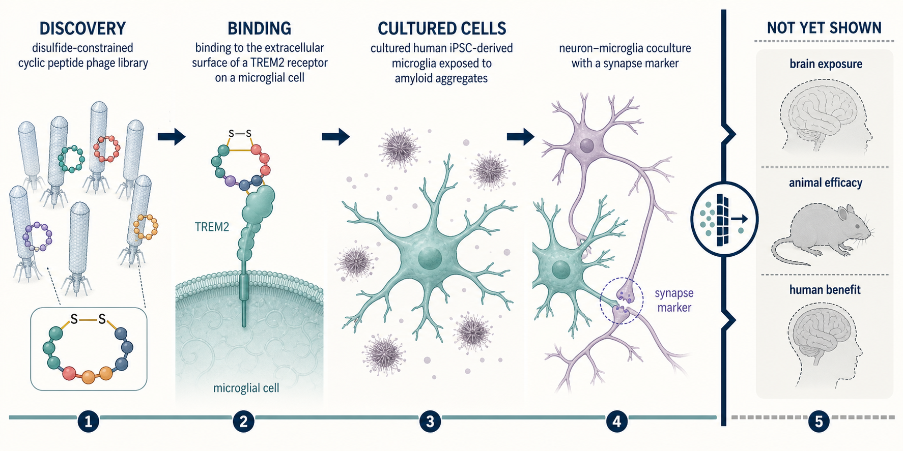
The study crossed discovery, binding, and cultured-cell gates. Brain exposure, animal efficacy, and human benefit remain untested.
What did the TREM2 cyclic peptide study find?
The researchers identified several TREM2-binding cyclic peptides, then advanced TREM2-6 and TREM2-12 because they combined the strongest measured binding with activity in cultured-cell models.
The discovery library used a CX9C format: nine variable amino acids sat between two cysteines, which form a disulfide bond and constrain the peptide into a loop. Recombinant human TREM2 extracellular domain was immobilized, and the library underwent four rounds of phage-display biopanning. The workflow enriched phage that remained associated with TREM2 after binding, washing, elution, and amplification.
Ninety-six clones were randomly sequenced in each of rounds 2, 3, and 4. Five recurring, sequence-diverse candidates were prioritized:
- TREM2-3:
CLPQQSQQTNC - TREM2-5:
CKKQWQKKARC - TREM2-6:
CRPAIAHIRTC - TREM2-10:
CENLNLYNMIC - TREM2-12:
CRKWAQNTE(Abu)C
For synthesized TREM2-12, 2-aminobutyric acid (Abu) replaced an internal cysteine from the phage-encoded sequence. That substitution left one unambiguous disulfide connection between the two flanking cysteines. The supporting information reports no conserved residue across the five sequences other than those flanking cysteines.
Phage ELISA showed preferential binding to TREM2-coated wells versus wells without protein, with three replicates. This is useful confirmation, but it is still an immobilized-protein assay—not proof that a free peptide binds the receptor on living brain cells.
How strong was the measured TREM2 binding?
The binding was real enough to prioritize leads, but it was micromolar rather than exceptionally tight.
Ten synthesized candidates were screened at 50 micromolar using a Monolith X spectral-shift assay. Assay buffer was tested in four negative-control replicates, while the previously reported TREM2 binder T2337 was used as a positive control in two replicates. Six candidates initially crossed the prespecified five-standard-deviation hit boundary. TREM2-11 was excluded because the instrument flagged aggregation and capillary adsorption.
Five remaining peptides passed dye-only interference controls and entered 16-point concentration-response testing beginning at 100 micromolar final concentration. Each affinity experiment was repeated independently three times. Reported dissociation constants were:
| Peptide | Measured KD (mean ± SEM) | |---|---:| | TREM2-3 | 48.3 ± 20.1 µM | | TREM2-5 | 41.7 ± 10.5 µM | | TREM2-6 | 16.4 ± 4.89 µM | | TREM2-10 | 57.8 ± 13.9 µM | | TREM2-12 | 10.2 ± 1.24 µM |
TREM2-12 therefore had the lowest reported KD, followed by TREM2-6. Lower KD generally means tighter binding under that assay’s conditions. It does not establish agonism, selectivity across the proteome, receptor occupancy in tissue, or useful exposure in a living organism.
What changed in the microglia experiments?
In amyloid-stressed cultured cells, TREM2-6 and TREM2-12 changed inflammatory, lipid-associated, and synaptic-marker readouts.
Human induced-pluripotent-stem-cell-derived microglia were pretreated for one hour with 5, 10, or 25 micromolar peptide, then exposed to amyloid-beta 1–42 oligomers. After 24 hours, the researchers measured IL-1β secretion. Both peptides reduced the amyloid-associated IL-1β signal relative to the amyloid-plus-vehicle condition, with TREM2-12 producing a modestly stronger pattern across the tested concentrations.
The cellular controls mattered:
- vehicle-treated cells with and without amyloid were included;
- VG-3927, a small-molecule TREM2 agonist, served as a positive control;
- wild-type and TREM2-knockout microglia were treated in parallel;
- the peptide-associated IL-1β effect was markedly attenuated in knockout cells, supporting TREM2 dependence;
- secreted ApoE was also measured after amyloid challenge and peptide treatment.
In a separate human iPSC-derived neuron–microglia coculture, the investigators used a 5:1 neuron-to-microglia ratio, exposed the cultures to amyloid, and measured PSD95, a postsynaptic protein used here as a synaptic-marker readout. Both peptides increased the reported “percent rescue” of PSD95 versus amyloid-treated vehicle controls.
The cell figures report mean ± SD with n = 5. One- or two-way ANOVA with Dunnett’s post-hoc test was used as appropriate, with significance set at p < 0.05. The methods also state that experiments had at least three independent replicates.
One reporting limitation deserves attention: the paper describes commercial human iPSC-derived microglia and neurons but does not identify the supplier, donor identities, number of distinct donor lines, sex, genotype, or differentiation batches. That makes the model difficult to interpret as evidence of reproducibility across genetically diverse people. Human-derived cells are not the same thing as a study in humans.
Did the study prove the peptides are TREM2 agonists?
Not fully. It supports TREM2-dependent functional modulation in these cell systems, but “agonist” is a stronger mechanistic label than the reported experiments conclusively establish.
The knockout experiment strengthens the causal link to TREM2. The concentration-related IL-1β response, ApoE change, and PSD95 result show that binding was accompanied by biological effects in the selected models. But the study did not report a complete receptor-signaling map, direct receptor-occupancy measurement, broad off-target panel, or independent replication in multiple donor backgrounds.
“Tune” or “modulate” is therefore the defensible description. Whether these peptides act as classical agonists, biased modulators, clustering agents, or through a more complicated receptor-context effect needs additional experiments.
What did molecular dynamics add?
The simulations supplied plausible binding models; they did not independently prove the physical binding site or mechanism.
The authors generated TREM2–peptide complexes with AlphaFold3 and ran 100-nanosecond molecular-dynamics simulations at 300 K. Both modeled complexes remained stable. TREM2-12 showed less conformational movement than TREM2-6, consistent with a more constrained modeled binding pose.
The models proposed distributed contacts on TREM2’s exposed surface rather than binding in a deep pocket. That is chemically plausible for cyclic peptides and broadly consistent with the phage-display result. Still, a stable computer simulation begins from a predicted structure. Structural methods such as crystallography, cryo-EM, NMR, or carefully designed mutational mapping would be needed to validate the binding pose experimentally.
What did the stability and permeability tests show?
The leads resisted degradation in plasma and simulated intestinal fluid better than in acidic gastric conditions, but both crossed a Caco-2 intestinal-cell layer poorly.
| In-vitro property | TREM2-6 | TREM2-12 | |---|---:|---:| | Simulated gastric-fluid half-life | 0.78 h | 0.89 h | | Simulated intestinal-fluid half-life | 4.90 h | 5.92 h | | Remaining in human plasma after 1 h | 74.6% | 79.5% | | Caco-2 permeability, A→B | 0.92 × 10⁻⁶ cm/s | 0.68 × 10⁻⁶ cm/s | | Rat-liver-microsome half-life | 2.35 h | 3.14 h |
The reverse-direction Caco-2 values were higher than the absorptive-direction values, which the authors interpreted as possible moderate efflux. These assays are useful early filters, but a Caco-2 monolayer models intestinal transport—not the blood–brain barrier. No brain-permeability assay, pharmacokinetic animal study, or measurement of peptide in brain tissue was reported.
Favorable stability does not rescue poor permeability. A molecule can survive in a test tube and still fail to reach its target in a living brain.
Why is this still a useful platform result?
The study shows that disulfide-constrained phage display can produce small, tunable ligands for a disease-relevant microglial receptor and connect binding to selected cell-model responses.
That matters because antibodies are not the only possible way to engage TREM2. Cyclic peptides occupy a different design space: they can present relatively large interaction surfaces while remaining smaller and chemically more adjustable than antibodies. The diverse sequences found here also suggest there may be more than one way to contact TREM2’s exposed extracellular surface.
But a useful platform result is not a treatment result. Compared with antibodies, these leads currently have much weaker binding than many engineered biologics and unresolved delivery. Antibodies bring their own challenges—including size, tissue penetration, manufacturing, and receptor-clustering behavior—but this study did not directly compare efficacy, selectivity, exposure, or safety between modalities. It therefore does not establish superiority to an antibody approach. For a chemistry-focused example of why ring closure can improve one assay without establishing a better medicine, see what a cyclic-peptide crosslinking study actually measured.
For a broader explanation of why target binding and biological effect are separate gates, see AI peptide design: why binding is not the same as biological effect. The study also fits the evidence ladder explained in What preclinical actually means in peptide research.
What remains unknown before this could matter for Alzheimer’s disease?
Nearly every therapeutic question remains open. The paper does not establish:
- oral absorption or any effective route of delivery;
- blood–brain barrier penetration or brain exposure;
- TREM2 target engagement in a living organism;
- pharmacokinetics or dose–exposure relationships in animals;
- activity in an animal model of amyloid pathology;
- cognitive benefit;
- modification of Alzheimer’s disease biology or progression;
- long-term immune, neurological, or systemic safety;
- selectivity against other receptors and proteins;
- reproducibility across multiple human donor lines;
- superiority to antibodies, small molecules, or other TREM2 strategies;
- safety or efficacy in people.
The published article states that the authors had no competing financial interest. The article and supporting information do not report a patent, dedicated funding statement, or a data-availability statement. The supporting PDF supplies sequencing counts, assay traces, binding curves, and chemical characterization, but it is not an openly described raw-data repository.
Bottom line
TREM2-6 and TREM2-12 are credible early research leads, not Alzheimer’s treatments. The strongest conclusion is that cyclic-peptide discovery produced micromolar TREM2 binders whose effects in amyloid-stressed human iPSC-derived cell models were partly supported by a TREM2-knockout control. The next decisive work is not more promotional language around “neuroprotection.” It is optimization, selectivity testing, measured exposure, experimental target engagement, animal efficacy, delivery, and safety.
A separate catestatin neurodegeneration study likewise reported signals across postmortem human tissue, cell cultures, and mouse models without establishing a treatment in people.
This article is for general education and evidence literacy. It does not provide diagnosis, treatment selection, dosing, administration, sourcing, purchasing advice, or individualized medical guidance.
Study citation
Fuchs N, Yuan S, Kuncewicz K, Elhamouly MA, El Gaamouch F, Gabr MT. Phage Display-Derived Cyclic Peptides Target TREM2 and Modulate Microglial Responses Under Amyloid Stress. ACS Chemical Neuroscience. 2026;17(16):3082–3093. doi:10.1021/acschemneuro.6c00304. Supporting information: doi:10.1021/acschemneuro.6c00304.s001.