Did the Peptide Cause the Effect? What a TFA Counterion Study Reveals
Seeing a biological effect after a peptide preparation is not the same as establishing that the peptide sequence caused it. A non-peptide component can contribute to the same outcome being attributed to the sequence. Tang and colleagues’ study of trifluoroacetate, or TFA, makes that distinction concrete: several unrelated short peptides produced similar cholesterol changes in mice, and follow-up comparisons implicated their associated counterion.
Published in Nature Communications on September 2, 2026, the paper offers an enduring lesson in TFA counterion peptide research: identify both the material being tested and what the comparison can actually establish.
Quick answer: TFA is a non-peptide counterion associated with some peptide salts—not a new therapeutic peptide. In this study, TFA alone reproduced lipid effects in certain mice, while one peptide lost its observed cholesterol-lowering effect when its TFA counterion was replaced. Responses varied by mouse strain, sex and endpoint. The animal studies were not blinded, and human liver-cell reporter activation did not reproduce the downstream rodent-cell changes. There was no human clinical trial.
This is evidence interpretation, not evidence to use TFA or peptides for cholesterol or any condition.
On this page
- What the research tests
- What was compared
- Model-specific findings
- The PPAR-alpha explanation
- Human-cell findings
- Scrambled controls
- Study-appraisal checklist
- Limits
- Sources
What does TFA counterion peptide research actually test?
It tests whether a non-peptide component of a preparation can contribute to the measured effect. A peptide is a chain of amino acids; its sequence describes their order. A counterion is a separate charged chemical species associated with oppositely charged sites on the peptide.
Trifluoroacetate is the negatively charged form associated with trifluoroacetic acid. Some synthetic peptides are obtained as TFA salts because of how they are manufactured and purified. TFA is not another amino acid in the sequence, and its presence as a counterion is not automatically evidence of adulteration.
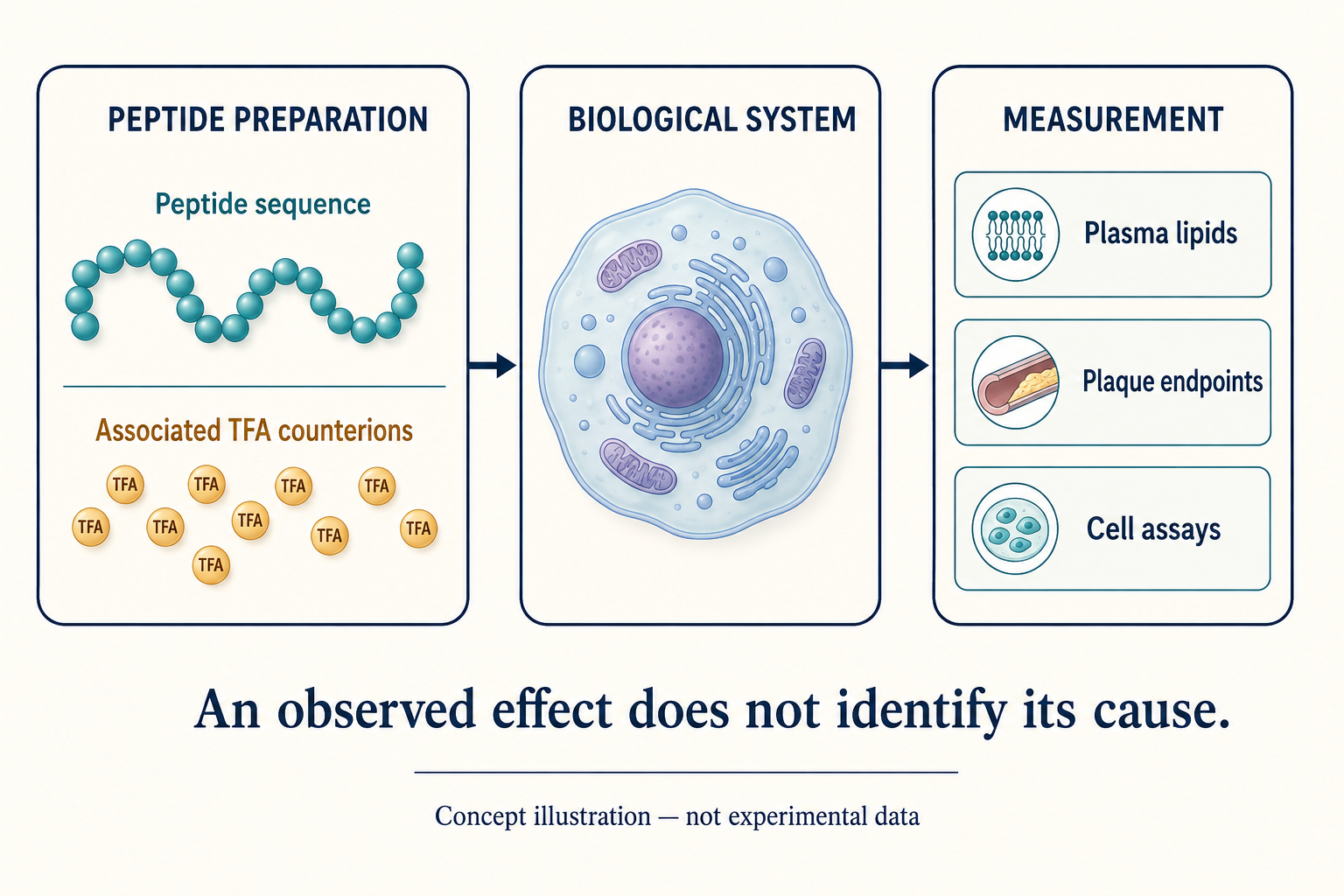
Original conceptual illustration, not experimental data. Arrows show study flow, not proof of a mechanism. The cell symbolizes a biological system; lipid and plaque endpoints came from animal studies, not from that illustrated cell. Shapes and counts do not represent a chemical structure or counterion ratio.
The central distinction is between sequence identity, preparation composition, and the outcome measured. Knowing the sequence does not fully characterize the material. Measuring an outcome does not identify its cause.
The same measurement discipline applies when a formulation property changes: our explainer on peptide hydrogel stability versus potency shows why improved stability alone does not establish preserved biological activity or clinical shelf life.
Counterion content can vary with the peptide’s charged sites and the particular preparation. Consequently, equal amounts of peptide material do not necessarily mean equal amounts of associated TFA. The broader characterization issue is explained in our synthetic-peptide manufacturing-quality guide; this article focuses on experimental attribution, not manufacturing standards.
What was actually compared?
The attribution argument came from several complementary comparisons, not merely from noticing that TFA was present. Each answered a different question.
Different sequences, similar unexpected effects
The researchers were investigating an earlier peptide finding when they tested four short fragments and two unrelated short peptides. These included a previously reported ineffective control. All six short peptide TFA salts reduced total plasma cholesterol relative to vehicle in female, high-fat-fed Ldlr-deficient mice. Reported reductions ranged from 24% to 55%.
That was a clue against a simple sequence-specific explanation. It was not, by itself, proof of the alternative cause: unrelated materials can share more than one property.
Figure 1’s cohorts were not uniformly eight mice. They were: vehicle n=23; parent peptide n=8; FREL n=4; KERS n=7; CGVLESF n=8; KASF n=6; EEWTKKLQ n=7; LSALEE-TFA n=9; and LSALEE chloride salt n=7. Here, n counts mice, not repeated measurements.
The figure used one-way analysis of variance (ANOVA) with Sidak-adjusted comparisons against vehicle. The six short-peptide TFA groups were statistically significant; the chloride-salt group was not. Those statistics compare each group with vehicle—not automatically every peptide directly with every other peptide.
The same sequence with a different counterion
For LSALEE, the authors compared TFA and chloride salt forms. Analytical testing confirmed TFA removal. The TFA form showed lower cholesterol than vehicle; the chloride form did not show the same effect.
This comparison strengthens the attribution to preparation composition while holding the sequence constant. However, “significant versus vehicle” in one group and “not significant versus vehicle” in another does not, on its own, supply a direct statistical test between those two groups. TFA-alone experiments help address that attribution gap.
TFA alone and a separate salt comparator
The authors then tested TFA without a peptide, using sodium trifluoroacetate. TFA alone reduced cholesterol under responsive Ldlr-deficient mouse conditions. Sodium acetate, included as a molar-matched comparator, did not reproduce that finding.
Together, the sequence comparisons, counterion-form comparison and TFA-alone results provide a stronger case than any one contrast alone. They show that the measured effect need not require the tested peptide sequence. They do not establish that every peptide has the same explanation.
Did all three mouse models show the same result?
No. Similar mechanistic signals did not produce uniform lipid or plaque outcomes. Plasma lipids are circulating measurements. Plaque endpoints assess lesions in arteries. Neither should be silently substituted for the other.
Ldlr-deficient mice: the clearest lipid pattern
Ldlr-deficient mice lack a working LDL receptor. In high-fat-fed animals, the study reported strong cholesterol and triglyceride reductions, with lipid effects reported in both sexes across experiments. Lipoprotein profiling supported reductions in the VLDL/LDL region rather than HDL. That profiling used pooled plasma from three mice per group, with the experiment repeated twice; it was not a separate large individual-animal LDL trial.
The longer plaque study used female mice only. After ten weeks, TFA-treated mice had 41% lower aortic-sinus lesion volume and 46% lower whole-aorta plaque area than vehicle controls. These were different anatomical measurements, not two names for one outcome.
Figure 2c reports n=8 TFA and n=7 vehicle, with unpaired two-tailed Student’s t-tests: p=0.0003 for sinus lesion volume and p=0.00001 for whole-aorta plaque area. These findings concern lesion development in female mice—not demonstrated reversal of established plaques or fewer human heart attacks.
The female high-fat triglyceride experiment in Figure 4b had n=16 vehicle, and n=7, 10 and 7 across its three TFA groups. The middle and highest groups were significant (p=0.0006 and p=0.007); the lowest was not, using ANOVA with Sidak comparisons. The corresponding liver-oxidation panel had only n=7 vehicle, illustrating why sample size must be checked by endpoint.
The result also had boundaries: male chow-fed Ldlr-deficient mice did not show the same cholesterol or triglyceride reductions, and a separate lower-exposure study did not show significant lipid or plaque changes. Diet and sex differed together in the Figure 4 Ldlr comparison, so that panel alone cannot separate their contributions.
Apoe-deficient mice: unchanged lipids, a male aortic plaque finding
Apoe-deficient mice did not reproduce the Ldlr lipid pattern. These animals lack apolipoprotein E, creating a different disturbance in lipoprotein handling.
The two-week lipid studies reported no significant cholesterol or triglyceride changes. Supplementary Figure 16 specifies n=7 per female group and n=8 per male group. Female comparisons used ANOVA with Sidak adjustment; male comparisons used unpaired two-tailed t-tests.
In the ten-week plaque experiment, male mice had 30% lower whole-aorta plaque area with TFA (p=0.02), but the 26% lower aortic-sinus lesion volume did not reach statistical significance (p=0.06). Both comparisons used n=9 vehicle and n=10 TFA, with ANOVA/Sidak testing reported in Figure 4e and Supplementary Figure 17.
Female plaque groups had n=10 each, with no significant benefit on these endpoints. A positive result in males and a non-significant result in females describes the observed pattern; it does not itself establish a formally tested treatment-by-sex interaction.
Liver fatty-acid oxidation increased in both sexes. That mechanistic finding does not turn the unchanged plasma lipids or non-significant plaque measurements into positive results.
Wild-type C57BL/6J mice: selected effects in males
Only males were studied in this metabolic-disease model. Triglycerides decreased significantly, but the lower cholesterol value was not statistically significant.
For Figure 4h, the high-fat comparison was n=14 vehicle versus n=10 TFA; a chow reference group had n=10. Triglycerides differed at p=0.004, using ANOVA with Sidak comparisons. Cholesterol was labelled non-significant; the figure does not provide an exact p-value for that contrast.
Liver palmitoyl-CoA oxidation increased (n=10 per group, Figure 4g), as did expression of two measured metabolic genes. These selected metabolic findings are not evidence of plaque reduction in wild-type mice or of the same response in females.
Endpoint check: “Changed liver metabolism,” “lowered circulating lipids,” and “reduced an artery-lesion measurement” are three different claims. Report the one actually supported in the relevant cohort.
How strong is the PPAR-alpha explanation?
Several assays support PPAR-alpha involvement, but its necessity for the mouse lipid and plaque effects was not established using knockout mice. PPAR-alpha is a gene-regulating protein involved in fat metabolism. Peroxisomes are small cellular compartments that help process fatty acids.
The supporting evidence included:
- Changes in mouse-liver genes associated with PPAR-alpha activity.
- Microscopy and protein measurements consistent with increased peroxisomes.
- Increased palmitoyl-CoA oxidation, a functional metabolic readout.
- Reporter assays indicating PPAR-alpha activation.
- A purified-protein coactivator-recruitment assay supporting a ligand-like interaction with PPAR-alpha.
There is an important distinction in the knockout evidence. The researchers did knock out PPAR-alpha in rat FAO liver cells. The tested gene-expression response to TFA was lost, supporting pathway dependence in that cell system (Figure 5a).
They did not perform the corresponding PPAR-alpha-knockout mouse confirmation. The discussion proposes those mice for future work, particularly to connect the mechanism to lipid changes. Rat-cell knockout evidence cannot by itself prove that this pathway explains every whole-animal endpoint.
Figure 5’s cell and binding results include technical triplicates and experiments repeated twice independently. Technical triplicates are repeated assay measurements, not three independent animals or three clinical participants.
What happened in human cells?
The human-cell findings did not reproduce the complete rodent response. TFA activated human PPAR-alpha in an engineered reporter assay in HepG2 cells, a human-derived liver cancer cell line. A reporter is a laboratory signal designed to reveal pathway activation.
But the authors reported neither the corresponding downstream gene-expression changes nor increased palmitoyl-CoA oxidation in TFA-treated HepG2 cells. Thus, pathway activation and the measured downstream metabolic response diverged in the human-derived cells.
A reporter lighting up is not equivalent to a beneficial change in liver metabolism, let alone lower cholesterol or better health in people. HepG2 experiments are cell research, not a human clinical trial. The study included no human participants. Our explainer on what preclinical actually means covers that boundary in more detail.
Does a shared counterion invalidate scrambled controls?
No—not automatically. A shared active component can complicate attribution without making every treatment-versus-control contrast meaningless. A scrambled peptide has its amino-acid order rearranged. That can test sequence specificity, but scrambling does not necessarily remove an associated counterion.
Consider two distinct comparisons:
- Peptide preparation versus vehicle: if the preparation includes TFA and vehicle does not, a difference cannot automatically be assigned to the peptide sequence.
- Peptide versus scrambled peptide, with verified equal counterion exposure: both groups may share a TFA-related effect, yet a difference between them can still provide evidence of a sequence-dependent contribution under those conditions, assuming other relevant factors are controlled.
Equal counterion content does not prove the shared component has no influence. It could interact differently with the preparations or affect the measurement’s responsiveness. But a shared component does not, by itself, explain away an observed between-group difference either.
Conversely, if both preparations differ from vehicle but not from each other, that comparison has not established sequence specificity. The reader needs the exact comparator, material characterization and counterion amount—not merely the word “control.”
This paper identifies a concrete attribution problem. It does not prove all synthetic-peptide results are artifacts, or invalidate any unrelated named peptide study without examining that study’s own materials and comparisons.
A reader’s study-appraisal checklist
Start with what differed between groups, then ask what the measurement can establish. These are questions for reading a paper, not instructions for conducting an experiment.
- What material was tested? Is the salt form and counterion content reported, or only the peptide sequence?
- What did the control contain? Vehicle alone, another peptide, the same sequence in another salt form, or the counterion alone?
- Was counterion exposure comparable? Equal peptide mass, sequence composition or a shared “TFA salt” label is not a measurement of equal TFA content.
- What was the endpoint? Lipids, plaque area, lesion volume, gene expression and reporter activity answer different questions.
- Which model and sex? Avoid combining female Ldlr plaque data, male Apoe plaque data and male wild-type triglyceride data into one universal effect.
- What does n count? Check animals, pooled samples, biological replicates and technical repeats separately.
- How was bias limited? Read allocation, blinding, exclusions and statistical comparisons—not just the abstract.
- Where does human relevance begin and end? Human-derived cells are not human clinical outcomes.
For the wider framework, see how to evaluate peptide claims online.
What are the study’s limits?
The study supports a specific counterion-attribution lesson, with important limits on precision and generalization. The counterion-form and TFA-alone comparisons strengthen attribution, while lack of blinding leaves a separate risk of bias.
The Methods state that animal studies were not blinded. Groups were described as randomized and balanced for age and weight, with different cage-allocation approaches for males and females. No data points were omitted. The reporting summary says no statistical method was used to determine sample size; the Methods describe typically n=8, chosen from prior experience—not a universal cohort size or a formal power calculation for every endpoint.
The many endpoints require attention to which comparison was tested and how multiple comparisons were handled. A small p-value does not identify a causal component by itself. A non-significant result does not prove exact equivalence or absence of any possible effect.
The paper is a peer-reviewed publisher Article in Press, not a preprint. Its findings do not establish human benefit, human safety, environmental-exposure effects, or a reason to start, stop or change a medicine.
The durable takeaway: ask whether the study demonstrated an effect of the preparation, a contribution from its counterion, or an effect attributable to its peptide sequence. Those are related—but different—conclusions.
Sources
The linked paper, full PDF, supplementary information and reporting summary support the study-specific statements above.
- Tang W and colleagues. Trifluoroacetate mediated reduction of atherosclerosis in mice. Nature Communications, September 2, 2026. DOI: 10.1038/s41467-026-77341-2.
- Full paper PDF: Figures 1–5; Results; Discussion; Methods, especially Animals and the PPAR-alpha cell-knockout section.
- Supplementary information: counterion characterization, lipoprotein profiling, null findings, cohort sizes and supporting cell assays.
- Reporting summary: sample-size rationale, randomization, lack of blinding, cell lines and absence of human participants.