Can a Self-Assembling Peptide Repair Tendons? What the Rat and Beagle Study Actually Shows
A self-assembling peptide called AsPep-FTSQ improved several measures of tendon structure and function in rats and beagles, but it has not been shown to repair tendons in people. The 2026 study is meaningful preclinical work: it links a specific biological mechanism to cell experiments and two animal species. It is not a human clinical trial, and it does not establish human safety, durable benefit, optimal administration, or superiority to rehabilitation or surgery.
The study, published in Nature Communications by Li and colleagues, tested a peptide designed to assemble into a local nanofibrous network and capture excess transforming growth factor beta 1 (TGF-β1). The results support further research into an engineered biomaterial. They do not validate consumer “healing peptide” claims.
Quick answer: The study shows that AsPep-FTSQ affected TGF-β1-related biology and improved selected imaging, tissue, and gait measures in surgically injured animal tendons. It does not show that the peptide repairs human tendons.
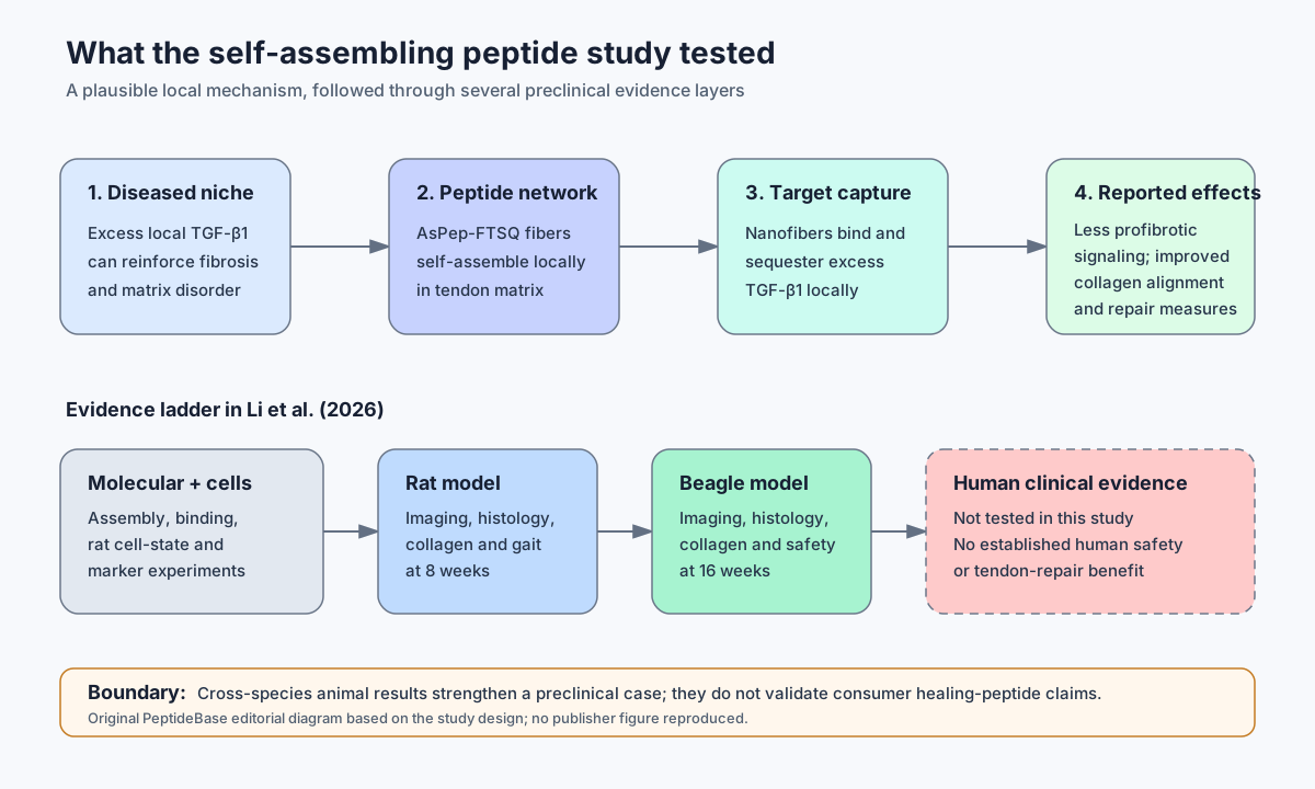 The paper connects a designed mechanism to cells, rats, and beagles. The missing step is the one that matters clinically: controlled human evidence.
What is the biological idea behind AsPep-FTSQ?
The premise is to reduce excessive local TGF-β1 activity without blocking the molecule throughout the body. TGF-β1 has normal roles in tissue repair and immune regulation, but persistently high signaling can also promote fibrosis, altered tendon-cell states, and disorganized extracellular matrix in tendinopathy.
AsPep-FTSQ was built from natural amino acids with two functional components. One portion supports self-assembly; another was selected to bind TGF-β1. In the researchers’ model, the peptide forms fibers around tendon cells and within tendon matrix, creating a local network that sequesters excess TGF-β1.
That distinction matters. The peptide was not presented as a generic growth signal or a replacement piece of tendon. It was designed as a local cytokine-capturing material intended to change the diseased tissue environment.
Mechanism is not outcome: Showing that a material binds a biological target makes the proposed explanation more plausible. It does not prove that the intervention is safe or useful in people.
What did the molecular and cell experiments show?
The early experiments supported both parts of the design: self-assembly and TGF-β1 capture. The authors used microscopy and biophysical testing to characterize nanofiber formation. Molecular simulations, surface plasmon resonance, and a competitive assay supported interaction with TGF-β1.
The cell work then modeled a fibrotic tendon environment by combining TGF-β1 exposure with cyclic mechanical stretch in primary rat tendon cells. Under those experimental conditions, AsPep-FTSQ was associated with lower expression of profibrotic markers and changes in tendon-related markers.
Single-cell RNA sequencing was used to characterize tendon-cell populations in the rat injury model. The analysis linked tendinopathy with shifts toward fibrotic, migratory, and osteo-chondrogenic cell states. The peptide experiments were consistent with limiting some of those pathological transitions.
These are useful mechanistic layers, but they have limits. The cell system used rat cells under constructed laboratory conditions. Single-cell analysis helps describe cell states and pathways; it does not by itself prove that one pathway causes a clinical outcome.
What happened in the rat tendon experiments?
In a surgically created rat Achilles tendon injury model, AsPep-FTSQ improved several structural, molecular, imaging, and gait-related measures at eight weeks. Rats were randomly assigned to four groups, with equal numbers of males and females. Histology scoring and imaging analyses were performed by investigators blinded to group allocation.
The comparisons included a sham group, the complete AsPep-FTSQ construct, and two partial peptide controls. The complete construct generally performed better than the control constructs. Reported findings included:
- lower tendon thickness and abnormal MRI signal
- improved ultrasound appearance and lower modified Ohberg scores
- lower Bonar histology scores
- less α-SMA and collagen I signal, alongside changes in tendon- and fibrosis-related genes
- more orderly collagen-fiber alignment
- less heterotopic mineralization on micro-CT
- improvements in paw contact, stride length, weight bearing, and Achilles functional index
Not every functional result changed. Swing-phase duration did not differ among groups. Most rat endpoint analyses used four animals per group; some molecular and functional analyses used three. Those small samples make estimates less stable, even when statistical tests produce low p-values.
The model is also important. Researchers partially transected Achilles tendon fibers and followed a controlled postoperative course. That produces a repeatable injury, but it is not the same as the varied overuse, metabolic, age-related, or degenerative tendon problems seen in humans.
What the rat study adds: It shows that the proposed mechanism was associated with whole-animal tissue and gait changes, not merely a marker change in cultured cells.
Did the beagle study replicate the result?
The beagle experiment reproduced several structural and molecular findings in a larger animal at 16 weeks, but it did not provide the same breadth of functional testing as the rat work. Beagles were randomly assigned to four groups, balanced by sex, and assessed with blinded histological and imaging analyses. Most reported comparisons used four animals per group.
Transmission electron microscopy showed a fibrous network within the tendon extracellular matrix that the authors interpreted as in-situ self-assembly. Compared with control peptide groups, the complete construct was associated with better gross tendon appearance, MRI measures closer to sham, lower Bonar scores, improved collagen alignment, and lower α-SMA and collagen I signals.
This cross-species replication is a real strength. Larger-animal anatomy and tissue loading can sometimes expose problems that rodent studies miss. But “worked in two species” is still not “works in humans.” The beagle study remained small, used an induced surgical model, and ended after four months.
What did the study report about safety?
The study found no obvious systemic toxicity signal in the tested beagles during four months, but it cannot establish human safety or detect uncommon harms. The researchers tracked blood counts and liver, kidney, and cardiac laboratory markers over time. At the endpoint, they examined the heart, liver, kidneys, lungs, and spleen by histology. They reported no treatment-specific laboratory abnormalities or major organ damage.
Rat body-weight trajectories also stayed within the expected range. These observations are reassuring within the experiment, but the boundary is narrow:
- four animals per group cannot reliably reveal rare adverse effects
- four months does not establish long-term durability or delayed toxicity
- normal systemic tests do not rule out subtle local tissue effects
- stage-dependent TGF-β1 biology may matter, because the pathway can support early repair as well as later fibrosis
- animal tolerability does not determine human safety
The authors themselves noted that future studies need to determine whether treatment timing and intensity should differ between acute and chronic stages.
How strong were the methods and statistics?
The design included useful safeguards, but the small samples and preclinical models limit certainty. Animals were randomized, both sexes were represented, and key imaging and histological assessments were blinded. The paper used partial peptide controls to test whether the full modular design mattered.
Results were generally reported as mean plus or minus standard deviation. Two-group comparisons used two-tailed unpaired Student’s t-tests; multi-group analyses used one- or two-way ANOVA with Dunnett or Tukey post-hoc testing. Statistical significance was set at p < 0.05. Cell and biochemical experiments were reported as at least three independent experiments.
Several cautions remain. The accepted manuscript does not describe a prospective power calculation. Many outcomes were tested across small groups, and the paper emphasizes p-values more than confidence intervals or prespecified primary endpoints. The single-cell differential-expression analysis was performed at the individual-cell level rather than with a pseudobulk approach, which can make biological replication and cell-level precision easy to confuse.
These issues do not erase the findings. They limit how precisely the findings should be interpreted.
Does this prove self-assembling peptide tendon regeneration in humans?
No. There were no human participants and no clinical outcomes. The study does not establish:
- whether AsPep-FTSQ is safe in people
- whether it improves pain, mobility, return to activity, or re-injury rates
- whether any effect lasts beyond the study windows
- which tendon conditions or disease stages might be relevant
- an optimal clinical formulation or administration strategy
- superiority or equivalence to progressive rehabilitation, surgery, or established care
Animal models reduce uncertainty; they do not remove the translation gap. Human tendinopathy varies by tendon, duration, loading history, age, health status, and prior treatment. A controlled surgical lesion in a healthy laboratory animal cannot reproduce all of that complexity.
Bottom line: This is credible preclinical evidence for a research platform, not clinical evidence for a tendon treatment.
Why this study does not validate BPC-157 or TB-500 claims
AsPep-FTSQ is a distinct engineered peptide material, so its results cannot be transferred to BPC-157, TB-500, or products marketed under those names. Different sequences, structures, targets, formulations, purity, and pharmacology mean different evidence requirements.
The study’s value comes from testing one defined construct against its proposed mechanism, partial controls, and animal endpoints. It does not support the broader idea that any peptide described as “healing” should repair connective tissue. That category leap is marketing, not inference.
Readers evaluating recovery claims can use the broader peptides for recovery evidence guide and the explainer on what preclinical actually means. The three-case nerve-injury report evidence guide shows why even observed improvement after BPC-157- or TB-500-containing multimodal treatment cannot isolate a peptide effect. For claim-checking beyond this study, see how to evaluate peptide claims online.
Funding, affiliations, patents, and conflicts
The work was conducted by academic hospital and university teams in China and was publicly funded. Authors were affiliated with Beijing Tsinghua Changgung Hospital/Tsinghua University, Peking University Third Hospital/Peking University Health Science Center, and Tongren Hospital/Shanghai Jiao Tong University School of Medicine.
Funding came from Chinese national, municipal, postdoctoral, clinical-translation, and research programs, including the National Natural Science Foundation of China and National Key Research and Development Program of China. The authors declared no competing interests. No patent disclosure or patent statement was identified in the paper or its declarations; that is not proof that no related intellectual property exists.
The article was published open access under a CC BY-NC-ND 4.0 licence. The study received animal ethics approvals from the Peking University Health Science Center committee.
The practical evidence-literacy takeaway
Li et al. provide a coherent preclinical chain: target biology, material design, binding and cell-state evidence, rat outcomes, and partial replication in beagles. That chain is stronger than a single in-vitro assay because multiple evidence layers point in the same direction.
The appropriate conclusion is still modest. AsPep-FTSQ deserves further toxicology, formulation, durability, and controlled human study. Until those steps happen, calling it a proven tendon repair treatment—or using it to advertise unrelated “healing peptides”—runs well past the evidence.
Frequently asked questions
What is AsPep-FTSQ?
AsPep-FTSQ is an experimental peptide made from natural amino acids and designed to self-assemble into nanofibers that bind excess TGF-β1 in local tendon tissue.
Was AsPep-FTSQ tested in humans?
No. The 2026 paper reported laboratory, rat, and beagle experiments. It was not a human clinical trial.
Did the animals show functional improvement?
Rats showed improvement in several gait and Achilles functional index measures, although swing time did not differ. The beagle work mainly reported imaging, histology, molecular markers, and systemic safety observations rather than a comparable functional battery.
Does natural-amino-acid design mean the peptide is safe?
No. Natural amino acids may simplify some design and manufacturing questions, but the complete assembled material still requires formal safety testing. Composition alone cannot establish safety.
Does this study support buying or using a tendon-healing peptide?
No. The research does not establish a consumer product, human treatment, dosing approach, or self-use case.
Source
Li C, Chen Z, Li W, et al. “A modular self-assembling peptide platform targeting TGF-β1 for tendon regeneration.” Nature Communications. Published August 11, 2026. DOI: 10.1038/s41467-026-76487-3.
This article is educational and is not medical advice. It does not recommend diagnosis, treatment, injection, dosing, purchasing, or changes to rehabilitation or clinical care.