Can a Self-Assembling Peptide Vaccine Make Cancer Immunotherapy Work Better? What the SaPAC Mouse Study Shows
SaPAC improved delivery, immune activation, and tumor control in mouse experiments, but it has not established human cancer-vaccine efficacy, safety, or personalized-treatment feasibility.
Can a Self-Assembling Peptide Vaccine Make Cancer Immunotherapy Work Better? What the SaPAC Mouse Study Shows
A self-assembling peptide cancer vaccine improved antigen delivery, immune activation, and tumor control in several mouse experiments, but it has not been tested in people. In Wu et al.’s 2026 Advanced Science paper, the SaPAC platform performed better than several component or physical-mixture controls and, in one breast-cancer model, worked best when combined with anti-PD-1. Those are encouraging preclinical findings—not evidence of human efficacy, safety, personalized-vaccine feasibility, or superiority to cancer vaccines already in clinical development.
SaPAC stands for Self-Assembling Peptide-Adjuvant Conjugate. The researchers chemically joined selected antigen epitopes to 1V209, a Toll-like receptor 7 (TLR7) agonist. The resulting conjugates formed positively charged nanoparticles roughly 100–200 nanometres across in hydrated measurements.
Quick answer: The study supports a coherent mouse-stage mechanism: covalent antigen-adjuvant coupling changed particle behavior, increased lymph-node exposure, improved antigen presentation, and strengthened antitumor immune responses. It does not show that SaPAC treats cancer in humans.
This article is educational only. It does not provide treatment, trial-enrollment, dosing, sourcing, purchasing, product, or investment guidance.
What did the researchers actually engineer?
SaPAC is a chemically defined research platform that places an antigen and an innate immune stimulant on the same self-assembling peptide construct. It is not one universal vaccine and it is not a marketed personalized treatment.
The core parts were:
- a lysine-rich, positively charged peptide region;
- cathepsin-sensitive SLVR linkers;
- a selected antigen epitope;
- designated lysines covalently linked to 1V209, a TLR7 agonist.
The paper distinguishes SaPN, the self-assembling peptide nanoparticle without covalently attached 1V209, from SaPAC, the corresponding peptide-adjuvant conjugate. That distinction created an important control: researchers could compare a covalent construct with the same broad ingredients merely mixed together.
The constructs were made by Fmoc solid-phase peptide synthesis, purified to greater than 95%, and checked by high-resolution mass spectrometry. Transmission electron microscopy assessed dry-state morphology; nanoparticle tracking analysis measured hydrated size; zeta-potential measurements assessed surface charge.
Supplementary Table S2 reported median hydrodynamic diameters of 169 ± 18 nm for SaPAC-OVA, 169 ± 4 nm for SaPAC-MB49, and 116–144 nm for the three SaPAC-4T1 constructs. Their zeta potentials were positive, ranging from +14.56 to +37.66 mV. These values came from three or five independent batches depending on the construct.
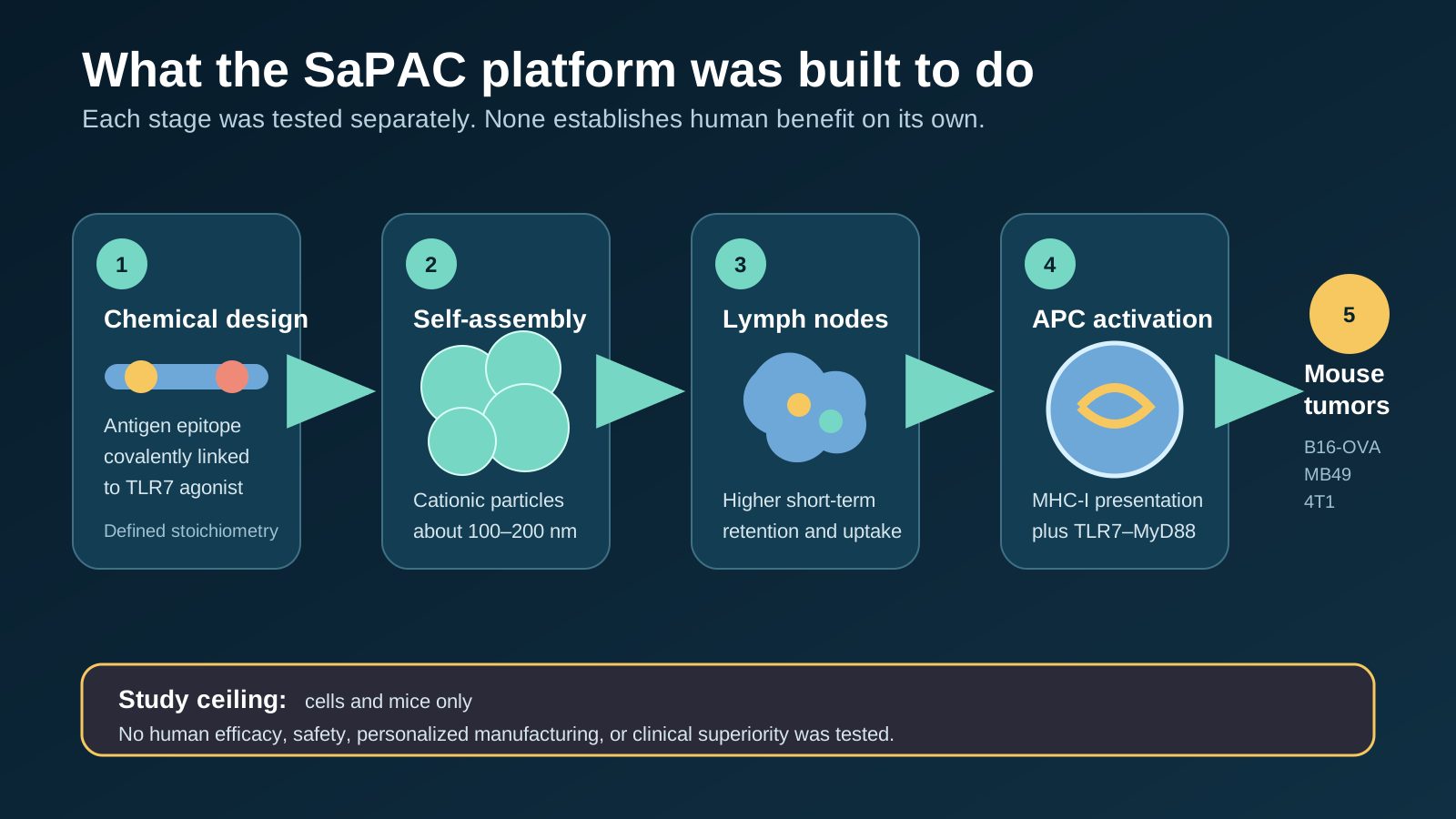
SaPAC links an antigen epitope and TLR7 agonist on one construct. Each arrow represents a separately tested evidence layer, not an automatic path to human benefit.
What did the lymphatic-drainage experiments show?
Fluorescent tracking suggested that the nanoparticle designs slowed rapid systemic escape and increased antigen exposure in draining lymph nodes. The first experiments focused on disposition, not tumor outcomes.
For the adjuvant-free precursor, FITC-SaPN-OVA produced stronger fluorescence in draining inguinal lymph nodes than soluble FITC-OVA six hours after subcutaneous injection. The paper reported p < 0.005. Pharmacokinetic fitting also indicated lower absorption and clearance rates for SaPN-OVA, while whole-body imaging showed longer retention near the injection site.
The SaPAC experiment then compared fluorescent Cy3-OVA, Cy3-SaPN-OVA, Cy3-SaPN-OVA mixed with free 1V209, and Cy3-SaPAC-OVA. At 24 hours, SaPAC-OVA showed the strongest reported signal in draining and distal lymph nodes and the highest intracellular signal in lymph-node dendritic cells. Supplementary Figure S6 specifies n = 4 mice per group for the organ biodistribution analysis.
The interpretation should stay narrow. Fluorescence is a proxy for labelled construct or label-associated material; it is not a direct measure of intact, functional vaccine over long periods. The paper’s own limitations note that lymph-node persistence was inferred mainly from short-term fluorescence imaging and pharmacokinetic modelling.
Evidence boundary: Better lymph-node exposure can make an immune mechanism more plausible. It does not establish tumor control, safety, or clinical value by itself.
Did SaPAC improve antigen-presenting-cell activation and MHC-I cross-presentation?
Yes, in cell experiments and short-term mouse immune assays, SaPAC produced stronger antigen-presentation and dendritic-cell activation signals than uncoupled controls. This is the strongest mechanistic part of the study.
In the DC2.4 mouse dendritic-cell line, approximately 90% of cells presented the OVA epitope after SaPAC-OVA exposure—nearly a 10-fold increase over SaPN-OVA or SaPN-OVA plus free 1V209. The authors also acknowledged that DC2.4 is a proliferative cell line with unusually high costimulatory-marker expression, which can complicate maturation readouts.
Primary bone-marrow-derived dendritic cells provided a more relevant follow-up:
- more than 75% of the SaPAC-OVA-treated CD11c-positive/MHC-II-positive cells displayed OVA through MHC class I;
- the corresponding values were below 50% for SaPN-OVA or SaPN-OVA plus free 1V209;
- more than 20% of OVA-presenting cells in the SaPAC group co-expressed the maturation markers CD80 and CD86—about four times the physical-mixture control.
In vivo, draining lymph nodes collected 24 hours after vaccination showed more mature dendritic cells and more CD69-positive activated T cells with SaPAC-OVA. The relevant figure reports n = 4–6 mice per group.
The proposed explanation is coordinated delivery. The linked construct brings antigen and 1V209 into the same cellular context; antigen can be processed for MHC-I cross-presentation while 1V209 activates endosomal TLR7 signalling through MyD88. A MyD88-null experiment supported that pathway: the enhanced IFN-gamma response was lost when MyD88 was absent.
What did the broader immune-cell readouts add?
The immune assays showed that SaPAC changed several steps between delivery and tumor growth, but they remained surrogate and mechanistic endpoints. They did not measure survival or patient benefit.
After prime-boost vaccination, SaPAC-OVA produced the highest reported frequency of IFN-gamma-secreting splenocytes and increased CD69 on CD8-positive and CD4-positive T cells after ex vivo restimulation. Cytokine profiling found higher TNF-alpha; Supplementary Figure S8 reports p = 0.0251 versus the stated comparator. IL-6 stayed near baseline relative to Poly(I:C)-adjuvanted groups.
Bulk RNA sequencing of draining lymph nodes identified 633 genes uniquely upregulated after SaPAC-OVA under the authors’ differential-expression thresholds. Pathway analysis highlighted TLR signalling, cytokine production, phagocytosis, and leukocyte activation. Single-sample enrichment suggested increased plasmacytoid dendritic-cell and macrophage signatures.
These analyses are supportive, not decisive. Gene-expression signatures infer pathway activity and cell-type enrichment; they do not count every recruited cell directly. Many immune outcomes were tested, increasing the importance of replication and transparent multiplicity control.
What happened in the three mouse tumor models?
SaPAC suppressed tumor growth in B16-OVA and MB49 experiments, while durable suppression in the 4T1 model required combination with anti-PD-1. The models answer different questions and should not be collapsed into one “cancer vaccine worked” claim.
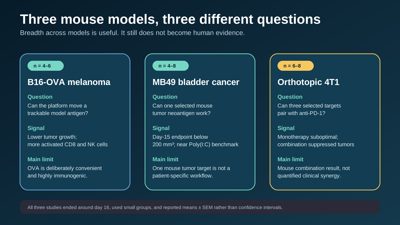
The three tumor systems increased biological breadth, but all remained short, small mouse experiments. OVA is a model antigen; MB49 and 4T1 used mouse tumor neoantigens.
B16-OVA melanoma: a deliberately immunogenic model-antigen test
SaPAC-OVA reduced tumor growth and was associated with more activated CD8-positive T cells and NK cells inside tumors. The longitudinal tumor figure reports n = 4–6 mice per group. Controls included OVA, SaPN-OVA, SaPN-OVA plus molar-matched free 1V209, and an OVA plus Poly(I:C) benchmark.
OVA means ovalbumin. The SIINFEKL epitope is widely used because mouse immune responses to it are easy to track. That makes B16-OVA useful for testing whether an engineered delivery system can move a known antigen through the immune pathway. It can also exaggerate how readily a real tumor antigen will be recognized.
Supplementary Figure S10 shows individual growth curves and notes that red crosses marked mice that died before the endpoint. The main figure and supplement therefore should not be read as if every animal contributed complete follow-up.
MB49 bladder carcinoma: one selected mouse tumor neoantigen
SaPAC-MB49 used a lab-screened MB49 neoantigen rather than OVA. Mice received four vaccinations, and the longitudinal figure reports n = 4–8 per group. SaPAC-MB49 reduced day-15 endpoint tumor volumes to less than 200 mm³, described as comparable to the peptide-plus-Poly(I:C) benchmark; unconjugated controls showed limited benefit.
This adds relevance because the target came from tumor mutation screening. It still does not reproduce the full problem of making a personalized vaccine for a person. The antigen was selected in one mouse tumor system, not prospectively manufactured for genetically diverse patients under clinical timelines and quality standards.
Orthotopic 4T1 breast cancer: combination therapy did the heavy lifting
Researchers screened candidate 4T1 mutations and selected three epitopes—4T1#2, #3, and #13—for a trivalent formulation. Supplementary Table S1 reports significant ELISPOT validation for each selected peptide (p < 0.0001); wild-type counterparts did not show significant cross-reactive immunogenicity in the reported screen.
As monotherapy, triSaPAC-4T1 produced suboptimal tumor suppression compared with the Poly(I:C) benchmark. In the combination experiment, anti-PD-1 alone had little activity, while triSaPAC-4T1 plus anti-PD-1 produced sustained suppression from day 9 onward. Endpoint volumes were described as comparable to the triple-neoantigen-plus-Poly(I:C)-plus-anti-PD-1 benchmark. The figure reports n = 6–8 mice per group.
That pattern is encouraging because it is consistent with a vaccine priming T cells that checkpoint blockade can help sustain. It does not prove clinical synergy. “Synergy” here describes the mouse combination result, not a quantified human interaction effect.
How large and certain were the reported effects?
Several mechanistic effects were large, but statistical reporting was less informative than the headlines sound. The paper commonly reported means ± SEM and significance thresholds from unpaired two-tailed Student’s t-tests.
The clearest numerical effects in the text were:
- about 90% antigen presentation in DC2.4 cells and a nearly 10-fold contrast with selected controls;
- more than 75% MHC-I presentation in primary dendritic cells versus less than 50% in selected controls;
- more than 20% mature antigen-presenting dendritic cells, about fourfold above the physical-mixture group;
- day-15 MB49 tumor volumes below 200 mm³ in the SaPAC group;
- particle sizes and charges reproduced across three to five independent batches.
For B16-OVA and 4T1 tumor growth, the article emphasized curves, endpoint comparisons, and significance symbols rather than reporting confidence intervals or a single prespecified effect estimate in the prose. The tumor groups were small and uneven in size. The many pairwise outcomes make isolated p-values easy to overread, and SEM describes precision around a sample mean rather than the spread among animals.
Quick answer: The results are compatible with a real platform effect in these experiments. Their precision, durability, and generalizability remain uncertain.
What did the study show about toxicity and safety?
The authors reported no detectable systemic toxicity in the tested mice, but this was not a formal human-safety assessment or a comprehensive translational toxicology program. The full paper and supplement do not turn that phrase into evidence about rare harms, delayed effects, immune-mediated toxicity, manufacturing impurities, or human TLR7 responses.
Short mouse studies can miss clinically important risks because group sizes are small, observation windows are brief, and species-specific immune signalling matters. Combination studies also cannot establish whether any future human toxicity would come from the vaccine, checkpoint blockade, their interaction, or a patient’s underlying disease.
The appropriate interpretation is “no systemic toxicity signal was detected under the reported mouse conditions,” not “the platform is safe.” The authors explicitly list formal safety testing, toxicology, GMP synthesis, dose-ranging, degradation, and batch comparability as work still needed for translation.
Does “precision” or “neoantigen” mean personalized medicine is ready?
No. The platform is modular, but modularity is not the same as a validated personalized-vaccine workflow. A clinically personalized product would require tumor and normal sequencing, mutation calling, expression and HLA-presentation prediction, antigen selection, rapid patient-specific manufacture, release testing, stability controls, regulatory review, and evidence that the resulting vaccine improves outcomes.
The mouse study demonstrated antigen swapping across OVA, one MB49 target, and three selected 4T1 targets. It did not test whether SaPAC can reliably complete that end-to-end process for patients, how often useful epitopes can be found, how quickly individualized lots can be produced, or whether the short selected peptides cover enough CD4-positive and CD8-positive responses across diverse HLA types.
It also did not compare SaPAC head-to-head with clinical personalized peptide vaccines or mRNA-LNP cancer vaccines in people. The paper discusses possible manufacturing and stoichiometric advantages, but those are proposed platform features—not demonstrated clinical superiority.
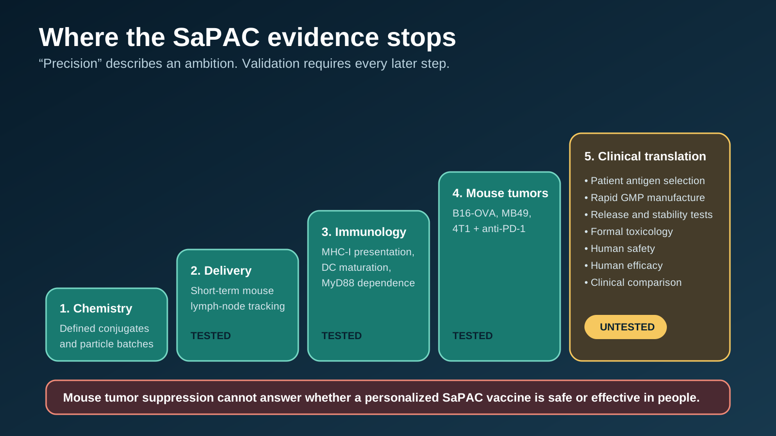
The study reached reproducible chemistry, immune mechanisms, and mouse tumor control. Human feasibility, safety, efficacy, and comparative value remain untested.
For the broader distinction between animal results and patient evidence, see What Preclinical Actually Means. For a human neoantigen-vaccine example, see An Immune Response Is Not a Survival Benefit. Our guide to evaluating peptide claims online applies the same evidence ladder to broader peptide marketing.
Who conducted and funded the research?
The author team spanned academic, hospital, government-research, and biotechnology affiliations, with disclosed commercial and patent interests. Affiliations included the University of Hong Kong’s biomedical sciences, biomedical engineering, cancer medicine, pathology, and pharmacology groups; Shenzhen Institutes of Advanced Technology; the University of Hong Kong-Shenzhen Hospital; Hong Kong Science and Technology Park; HKU-SIRI; BayVax Biotech Limited; and Shenzhen BayVax Biotech Limited.
The study reported support from Chinese national and Shenzhen research programs, HKU funds, the Innovation and Technology Support Programme, and related public or institutional sources.
The conflict statement says Ye-Fan Hu is CEO of BayVax Biotech Limited. It also says Jian-Dong Huang and Ye-Fan Hu are named inventors on a patent application related to the work, assigned to Versitech Limited and published as US 2025/0049902 A1, “Method of Developing a Peptide-Based Vaccine Conjugated with 1V209.” The author-contribution statement additionally disclosed use of Gemini 3.1, Grok 4, and DeepSeek V2 in writing, schematic diagrams, and analysis.
These disclosures do not invalidate the experiments. They increase the importance of independent replication, complete reporting, and clinical testing by groups beyond the platform’s inventors and commercial affiliates.
Bottom line
The SaPAC study is a well-layered preclinical platform paper, not a cancer-treatment result. It links defined chemistry to nanoparticle characterization, lymph-node behavior, dendritic-cell cross-presentation, TLR7-MyD88 signalling, immune activation, and tumor suppression in three mouse systems. Covalent coupling often outperformed simply mixing the peptide nanoparticle with free 1V209, which supports the authors’ central design claim.
The ceiling is equally clear. Small mouse groups, short follow-up, model-specific antigens, limited toxicity assessment, no human participants, no clinical manufacturing test, and no comparison with validated human cancer-vaccine platforms prevent conclusions about efficacy, safety, personalization, or superiority.
Frequently asked questions
What is SaPAC?
SaPAC is an experimental Self-Assembling Peptide-Adjuvant Conjugate platform. It covalently links an antigen-containing peptide construct to the TLR7 agonist 1V209 and self-assembles into cationic nanoparticles.
Is SaPAC a self-assembling peptide cancer vaccine?
It is a preclinical self-assembling peptide cancer-vaccine platform tested in cells and mice. It is not an approved vaccine or clinically validated treatment.
Was SaPAC tested in people?
No. The reported experiments used cell systems and mouse models.
What is OVA, and why does it matter?
OVA is ovalbumin, a model antigen. Its SIINFEKL epitope is easy to track in mice, making it useful for mechanism experiments but more immunologically convenient than many naturally occurring human tumor targets.
Did SaPAC work with anti-PD-1?
In the 4T1 mouse model, triSaPAC-4T1 plus anti-PD-1 suppressed tumors more durably than either component alone and performed comparably to a Poly(I:C)-adjuvanted benchmark combination. That does not establish clinical synergy.
Did the study prove SaPAC is safe?
No. The authors reported no detectable systemic toxicity under the tested mouse conditions, but formal translational toxicology and human safety remain unestablished.
Is SaPAC already a personalized cancer vaccine?
No. The study demonstrated modular antigen substitution in mouse systems. It did not validate patient-specific antigen selection, manufacturing, quality control, safety, or clinical benefit.
Sources
- Wu Y-F, Hu J-C, Hu Y-F, et al. “Self-Assembling Peptide-Adjuvant Conjugate (SaPAC) Platform for Precision Cancer Immunotherapy.” Advanced Science. Published October 2, 2026. doi:10.1002/advs.77632.
- PubMed Central. Full text, PMCID PMC13633104.
- PubMed. PMID 42827008.
- Wu et al. Supporting File, including Tables S1–S2 and Figures S1–S14. PMC Open Access dataset copy.
- NCBI BioProject. PRJNA1519071.