Can MOTS-c Protect the Diabetic Retina? What the New Cell and Mouse Study Shows
The short answer: A 2026 study found that MOTS-c was linked to better endothelial-barrier and mitochondrial measurements in cultured human retinal endothelial cells and in diabetic mice. The work also implicated PLA2G6 as a mediator inside those models. It did not test whether MOTS-c prevents blindness, treats diabetic retinopathy in people, or is safe and effective for human use.
Published online in Cell Death Discovery on October 10, 2026, the study is useful because it connects several evidence layers: where MOTS-c appears in the retina, what high glucose does to its expression, what happened when researchers added MOTS-c to cultured cells, what happened in a diabetic mouse model, and whether disrupting PLA2G6 weakened those effects.
That chain is more informative than a single pathway experiment. It is still a preclinical chain. Readers new to that distinction may want our guide to what preclinical evidence actually means and the broader MOTS-c evidence overview.
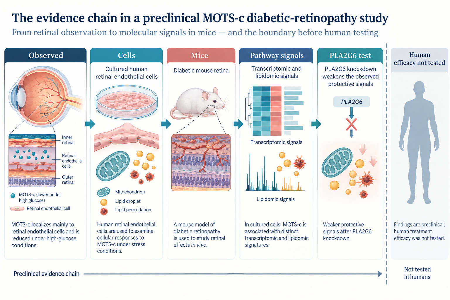
The paper builds a preclinical evidence chain across observations, cultured cells, mice, omics, and PLA2G6 knockdown. It stops before human treatment efficacy or safety.
What did the MOTS-c diabetic retinopathy study actually find?
The study found model-level protective signals, not a demonstrated treatment. Its main findings were:
- MOTS-c staining and mitochondrial transcript signals were concentrated in retinal vascular endothelial cells in the datasets and tissue analyses used by the authors.
- High-glucose conditions reduced MOTS-c-related RNA, intracellular protein, and secreted peptide measurements in cultured human retinal endothelial cells.
- Vitreous samples from 11 people with diabetic retinopathy had lower measured MOTS-c than samples from 10 nondiabetic controls undergoing surgery for macular hole or epiretinal membrane.
- Added MOTS-c improved several barrier, oxidative-stress, and mitochondrial readouts in high-glucose-exposed cells.
- In diabetic mice, MOTS-c was associated with less retinal vascular leakage, fewer acellular capillaries, better junction-protein measurements, and partial preservation of retinal structure and mitochondrial markers.
- Transcriptomic and lipidomic analyses pointed toward phospholipid metabolism, with PLA2G6 emerging as the leading candidate among the phospholipase-related genes examined.
- Knocking down PLA2G6 weakened many of the MOTS-c-associated signals in both cells and mice.
Those results support a mechanistic hypothesis: in these models, MOTS-c may help preserve retinal endothelial function partly through PLA2G6-linked handling of oxidized phospholipids. They do not establish that the same chain produces a meaningful visual benefit in people.
What was observed before MOTS-c was tested as an intervention?
The first evidence layer was localization and association. The researchers reported that MOTS-c was prominent in the vascular-rich inner retina and that its expression was enriched in endothelial-cell clusters in a public single-cell dataset. In cultured human retinal endothelial cells, exposure to 30 mM glucose for 48 hours reduced mitochondrial 12S-rRNA transcription, intracellular MOTS-c protein, and MOTS-c measured in the culture medium.
The supplement added an osmotic comparison: 5 mM glucose plus 25 mM mannitol did not reproduce the reduction seen with 30 mM glucose. That supports a glucose-related interpretation, but the authors correctly noted that the main experiments did not consistently include osmotic controls. Subtle osmotic effects therefore cannot be fully excluded.
The small human vitreous comparison adds context, not treatment evidence. The diabetic-retinopathy group contained 11 samples and the control group 10. The groups were similar in sex distribution but differed substantially in age: the diabetic-retinopathy group averaged 60.0 years and controls 74.2 years. These were surgical samples from different eye conditions, so lower vitreous MOTS-c is an association vulnerable to confounding. No participant received MOTS-c.
What happened in cultured human retinal endothelial cells?
MOTS-c improved several laboratory readouts under high-glucose stress. The investigators used authenticated primary human retinal endothelial cells from passages 1 through 6. Cells were compared under normal glucose, high glucose, high glucose plus MOTS-c, and several mechanistic control conditions.
The authors selected 5 micromolar MOTS-c for later cell experiments after a viability screen. Under high glucose, the peptide was associated with:
- lower mitochondrial superoxide and intracellular reactive-oxygen measurements;
- higher levels of junction proteins including ZO-1, occludin, and VE-cadherin;
- less internalization of VE-cadherin and beta-catenin;
- less FITC-dextran movement across a cultured endothelial layer;
- improved mitochondrial membrane potential, morphology, mitochondrial-DNA copy number, and several mitochondrial quality-control markers.
Actinonin, used as a functional way to suppress translation from mitochondrial 12S rRNA, generally pushed results in the opposite direction. But actinonin is not a MOTS-c-specific neutralizing tool. The authors acknowledged that a mitochondria-targeted RNA-interference method or validated neutralizing antibody would provide stronger specificity.
These are cellular stress and barrier assays. They can reveal a plausible mechanism, but a dish does not reproduce the intact retina, immune system, circulation, drug distribution, or clinical course of diabetic retinopathy.
What happened in the diabetic mice?
The mouse experiments produced retinal protection signals across several surrogate endpoints. Male C57BL/6J mice were placed on a high-fat diet and given streptozotocin to create a hyperglycemic diabetes model. Diabetic animals were compared with nondiabetic controls and diabetic animals receiving MOTS-c.
In the first experiment, the study schematic reports intraperitoneal MOTS-c at 5 mg/kg per day for seven days beginning seven weeks after diabetes onset, with tissue collection at week 12. In the PLA2G6 experiment, an endothelial-targeted AAV carrying either control shRNA or shPLA2G6 was delivered before diabetes induction; each eye later received 5 pmol of MOTS-c or vehicle at weeks 8 and 9, with collection at week 12.
Those details describe what the researchers did in mice. They are not a human protocol, dose conversion, or treatment instruction.
Across the main mouse figures, many histology and retinal endpoints appear to use five animals per group, while several protein-blot analyses show three observations per group. The paper says data came from at least three independent experiments unless otherwise stated, but it does not provide one consolidated sample-size table or a formal power calculation. The supplement says animals were computer-randomized and images were analyzed by a fully blinded investigator.
Reported outcomes included less Evans-blue leakage, fewer acellular capillaries, higher junction-protein levels, more preserved retinal thickness and NeuN-positive retinal ganglion-cell counts, and improved mitochondrial markers. These are relevant disease-model measurements. The study did not measure human vision, progression to blindness, or patient-important outcomes.
The model also has boundaries. It used only young male mice, combined high-fat feeding with streptozotocin, and examined a relatively early experimental window. Human diabetic retinopathy develops across years and includes substantial metabolic, vascular, neural, inflammatory, treatment, and demographic variation.
What did transcriptomics and lipidomics add?
The omics results generated a pathway hypothesis; they did not prove the pathway by themselves. RNA sequencing compared high-glucose-exposed cells treated with MOTS-c or vehicle, with three biological samples per group. Gene-set analyses highlighted phospholipase A2 activity, phosphatidylcholine catabolism, lysophospholipase activity, and other lipid-metabolism pathways.
Lipidomics used six samples per condition. MOTS-c-treated cells showed a different glycerophospholipid pattern, including higher lysophosphatidylethanolamine and bis-methyl phosphatidic acid, and lower phosphatidylinositol and phosphatidylserine. Because these were discovery-scale measurements with many genes and lipids, individual nominal P values deserve caution. The transcriptomic enrichment analysis used a false-discovery-rate threshold, but the lipidomics selection combined variable-importance scores with Student's t-tests, and the article did not center confidence intervals or effect-size precision.
The raw RNA-sequencing and lipidomic datasets were deposited on Figshare, which improves transparency. Still, omics signatures are best read as maps pointing toward follow-up experiments—not as clinical biomarkers or treatment outcomes.
Is PLA2G6 a demonstrated mediator or a clinically validated target?
PLA2G6 is a reasonably supported mediator within these experiments, but it is not a clinically validated diabetic-retinopathy target. PLA2G6 encodes a calcium-independent phospholipase involved in phospholipid remodeling. In the study, its expression fell under high glucose and in diabetic mouse retinas, then rose with MOTS-c treatment.
The researchers went beyond correlation in several ways:
- PLA2G6 was the strongest upregulated candidate among the phospholipase-related genes highlighted by the RNA analysis.
- Cell experiments used PLA2G6 siRNA, a pharmacologic inhibitor, and a pharmacologic activator.
- PLA2G6 knockdown weakened MOTS-c-associated improvements in lipid-peroxidation, mitochondrial, and endothelial-junction measurements.
- Endothelial-targeted Pla2g6 knockdown in mice weakened several retinal effects associated with MOTS-c.
That pattern supports mediation: when PLA2G6 activity or expression was reduced, much of the observed MOTS-c effect was reduced too. It does not prove that PLA2G6 is the only mediator, that MOTS-c directly binds or activates PLA2G6, or that manipulating this enzyme would be beneficial and safe in humans.
The authors explicitly noted that other phospholipase A2 family members were not systematically excluded and that the precise way MOTS-c activates PLA2G6 remains unknown. A target can be experimentally useful without being clinically validated. Drug development has buried entire graveyards of targets that looked terrific in mice. Biology is generous with clues and stingy with approvals.
How certain are the statistics?
The results are internally consistent, but precision and reproducibility remain uncertain. The paper generally reports means with standard deviations, uses t-tests for two groups and one-way ANOVA with post-hoc testing for multiple groups, and marks thresholded P values. Several limitations affect confidence:
- small assay-level sample sizes, often three to six;
- no reported prospective power calculation;
- many related molecular and imaging endpoints;
- limited reporting of confidence intervals and standardized effect sizes;
- biological-replicate dots mixed with individual-cell measurements in some figure captions;
- unclear independence when multiple images or cells contribute to an assay;
- no independent replication laboratory;
- no female mice and no human intervention cohort.
Statistical significance means the observed group difference was unlikely under the test's assumptions. It does not tell us that the effect is large enough to matter clinically, will reproduce, or will survive translation from cells and mice to people.
What are the study's main limitations?
The largest limitation is translation: no human being was treated. Other important limits include the small and age-imbalanced vitreous comparison, absence of osmotic controls from the main high-glucose series, imperfect specificity of actinonin, incomplete exclusion of other phospholipase-family pathways, and an unknown direct link between MOTS-c and PLA2G6 activation.
The mouse model is useful for controlled mechanistic work but does not recreate the full chronic human disease. Outcomes centered on vascular permeability, retinal structure, protein markers, oxidative stress, and mitochondrial biology—not visual acuity, visual-field preservation, need for eye procedures, or prevention of blindness.
Safety was also not established for human retinal or systemic use. Model tolerability and cell viability cannot substitute for clinical pharmacokinetics, immunogenicity, ocular safety, systemic adverse-event monitoring, or longer-term follow-up.
Who funded the study, and were conflicts disclosed?
The paper reports public and academic grant support and no competing interests. Funding came from the Basic Research Program of Jiangsu, the China Postdoctoral Science Foundation, the Jiangsu Funding Program for Excellent Postdoctoral Talent, and an open research project at the Nanjing University Institute of Traditional Chinese Medicine Integrative Chinese and Western Medicine Research program.
The authors declared no competing interests. The animal work had institutional ethics approval. The supplement says the vitreous repository and ELISA analysis received ethics approval and participants gave informed consent.
Funding and conflict statements are context, not a quality score. The more important questions remain whether the design fits the claim, whether the controls are adequate, whether the statistics are robust, and whether independent groups reproduce the result.
Bottom line: can MOTS-c protect the diabetic retina?
In cells and diabetic mice, the study reports several signals consistent with retinal endothelial and mitochondrial protection. In humans, the answer is unknown. PLA2G6 appears to mediate part of the effect inside the tested models, but it is not yet a validated clinical target for diabetic retinopathy.
The right next steps would include independent replication, stronger target-specific experiments, broader animal designs, pharmacology and safety work, and eventually carefully controlled human studies with clinically meaningful retinal and vision outcomes. Until then, this is promising preclinical biology—not proof of a treatment.
For another example of why cell and mouse findings must stay inside their evidence boundary, see our review of a MOTS-c host-defense study.
Frequently asked questions
Did the study show that MOTS-c prevents blindness?
No. It measured cellular, molecular, vascular, and retinal-tissue outcomes in cultured cells and mice. It did not test blindness prevention or visual outcomes in people.
Was MOTS-c tested in people with diabetic retinopathy?
No one received MOTS-c. The only human component was a small observational comparison of vitreous MOTS-c levels in surgical samples from 11 people with diabetic retinopathy and 10 controls.
Does PLA2G6 prove how MOTS-c works?
It supports one important mechanism in these models. Knockdown weakened multiple MOTS-c-associated effects, but other pathways were not fully excluded and the direct activation mechanism remains unresolved.
Does this paper establish a safe dose?
No. The cell and mouse exposures are experimental methods, not human dosing guidance. Human efficacy, ocular safety, systemic safety, and long-term risk were not tested.
Primary source
- Liao Y, Xiang J, Xia Y, et al. Mitochondrial-derived peptide MOTS-c rescues vascular endothelial damage via PLA2G6 in diabetic retinopathy. Cell Death Discovery. Article in Press, 2026. The full paper and supplementary methods, figures, and tables were reviewed for this article.
- Public study datasets: RNA sequencing and lipidomic profiling.
This article is for education and evidence interpretation only. It does not provide medical advice, dosing, treatment instructions, purchasing guidance, or sourcing advice.