Can AI Design Mirror-Image Peptides? What Mirror-Peptidizer Actually Proved
Yes—but “design” means generating and prioritizing candidates for experiments, not proving that those candidates are medicines. In an August 31, 2026 paper in Research, Ma and colleagues introduced Mirror-Peptidizer, an open-source workflow for creating AI-designed D-peptide candidates from an ordinary L-protein structure. The team then synthesized selected candidates and reported target binding or cell-based activity across MDM2, PD-L1, and IL-23R.
The experimental results make this more than a docking-only paper. Four of nine MDM2 candidates showed measurable binding, and the strongest, MBDP-1, had a reported dissociation constant (Kd) of 11.9 nM by isothermal titration calorimetry. But the study did not test animals or people. It did not establish pharmacokinetics, delivery in a living organism, toxicity, human efficacy, or clinical usefulness.
Quick answer: Mirror-Peptidizer proved that a mirror-coordinate computational workflow can produce D-peptide candidates that survive several laboratory tests. It did not prove that AI can design a safe or effective D-peptide drug on demand.
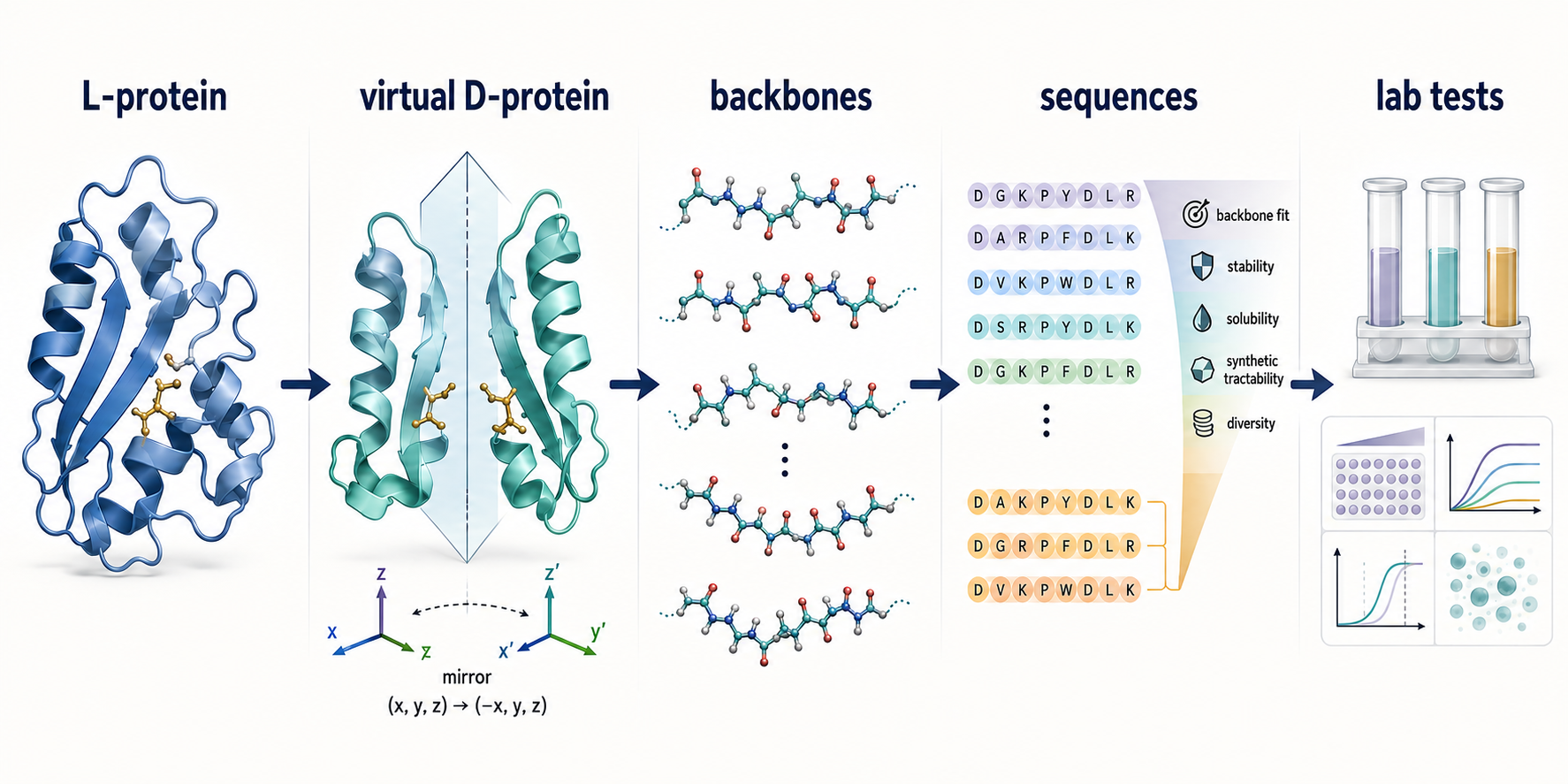
Mirror-Peptidizer narrows a large computational search to candidates worth testing. The ranking step uses sequence–backbone compatibility and tractability-related heuristics; binding is established only in later experiments.
What are D-peptides?
D-peptides are peptides built from D-amino acids, whose three-dimensional handedness is opposite to the L-amino acids that dominate ordinary proteins. A D-peptide can be thought of as the mirror image of its all-L counterpart, much as left and right hands contain corresponding parts but cannot be perfectly superimposed.
Handedness matters because biological recognition is three-dimensional. A peptide with the correct sequence but the wrong stereochemistry may fold differently or fail to fit a target. The reversed configuration can also make D-peptides less susceptible to many proteases, because those enzymes evolved mainly to recognize L-peptide substrates.
That potential protease resistance is useful, but it is not a complete drug profile. It does not automatically provide target selectivity, cell entry, tissue delivery, acceptable clearance, low toxicity, low immunogenicity, or clinical benefit.
Why has mirror-image discovery often required a synthetic D-protein?
Traditional mirror-image phage display solves the handedness problem experimentally, but it usually requires a chemically synthesized mirror version of the target protein. Researchers screen an L-peptide library against that D-protein target. If an L-peptide binds the mirror target, converting the peptide to its D-form should create a corresponding binder for the natural L-protein.
The logic is elegant:
- Build a D-form version of the target protein.
- Screen ordinary L-peptides against that mirror target.
- Identify an L-peptide binder.
- Convert the hit to the matching D-peptide for the native L-target.
The bottleneck is step one. Chemically producing a correctly folded D-protein can be labor-intensive, especially for larger or structurally complicated targets. Mirror-Peptidizer tries to replace the physical mirror target during candidate generation with a virtual one.
How does Mirror-Peptidizer design AI-generated D-peptides?
Mirror-Peptidizer works in a computational L-amino-acid design space, then mirrors the result back into a D-peptide predicted to fit the native target. It does not ask a general model to invent a drug in one step.
1. Mirror the target coordinates
The workflow begins with a three-dimensional structure of the native L-protein. It inverts one coordinate axis to create a virtual D-protein. The paper reports chirality checks showing the expected mirror relationship across paired backbone dihedral angles, while explicitly noting that Chroma and ProteinMPNN are not themselves chirality-aware models.
2. Generate candidate backbones with Chroma
Chroma, a diffusion-based protein design system, generates L-peptide backbone shapes against the fixed virtual D-target. Geometry filters remove poses that are buried, badly positioned, distorted, or implausibly connected to the target surface.
3. Assign sequences with ProteinMPNN
ProteinMPNN proposes amino-acid sequences compatible with each generated backbone while the receptor remains fixed. Its score is a measure of sequence–backbone compatibility. It is not an experimental affinity measurement.
4. Explore nearby sequences with Bayesian optimization
Bayesian optimization makes limited sequence changes and balances two main priorities: the ProteinMPNN compatibility score and a solubility or synthesis-related heuristic. The authors combine normalized desirability values into a FuzzyScore so a severe weakness in one objective can drag down the overall rank.
5. Mirror and test the prioritized candidates
The selected L-peptide–virtual-D-target complexes are mirrored back, producing D-peptide candidates for the native L-protein. Only then are selected molecules synthesized and tested.
Critical distinction: The pipeline ranks candidates; it does not explicitly calculate the final intermolecular binding energy. The authors say the top-ranked sequences should be interpreted as experimentally testable priorities, not globally optimal binders.
What did the MDM2 experiments prove?
The MDM2 series provided the paper’s strongest binding result and its most detailed interface validation, but five of nine synthesized candidates did not show measurable binding. That failure rate is important evidence about what the model can and cannot guarantee.
The team synthesized nine prioritized MDM2-binding D-peptides, called MBDPs, and tested them by isothermal titration calorimetry (ITC):
| Candidate | Reported ITC Kd | Interpretation | |---|---:|---| | MBDP-1 | 11.9 nM | Strongest reported MDM2 binder | | MBDP-2 | 86 nM | Nanomolar binding | | MBDP-3 | 584 nM | Sub-micromolar binding | | MBDP-4 | 876 nM | Sub-micromolar binding | | Remaining five | No measurable affinity reported | Computational prioritization did not ensure binding |
An orthogonal biolayer interferometry measurement put MBDP-1 at 29 nM, broadly consistent with the ITC result. Nuclear magnetic resonance chemical-shift changes placed MBDP-1 in the p53-binding region of MDM2. Mutating predicted contact residues also weakened binding: Y100A shifted the reported Kd to 632 nM, F55A to 233 nM, and K51A to 30.3 nM. Together, these tests support the proposed interface more strongly than a single affinity trace would.
The study also attached a six-arginine cell-penetrating sequence to selected MBDPs and tested HCT116 colorectal cancer cells. The conjugates reduced viability in p53-positive cells but not p53-knockout cells, and the authors reported apoptosis and increased p53 and p21 signals. R6 alone and weak or inactive peptide conjugates served as additional controls.
Those are mechanistically informative cell experiments. They are not animal efficacy, tumor response in a living organism, systemic delivery, or evidence of safety in humans.
What did the PD-L1 and IL-23R tests add?
The PD-L1 and IL-23R results suggest that the workflow can generate different peptide shapes for different target surfaces, but the experimental funnels were smaller than the full computational search. The reported conformations included a beta-rich PD-L1 binder and a mixed helical/flexible IL-23R binder, alongside the more helical MDM2 series.
| Target | Candidates experimentally highlighted | Binding evidence | Functional evidence | |---|---|---|---| | MDM2 | 9 synthesized; 4 bound | MBDP-1 ITC Kd 11.9 nM; BLI Kd 29 nM; NMR and mutant controls | Cell-penetrating conjugates produced p53-dependent effects in HCT116 cells | | PD-L1 | 4 synthesized; 2 bound | PBDP-1 ITC Kd 64.9 nM; PBDP-2 313 nM; PBDP-3 and -4 showed no measurable affinity | PBDP-1 and -2 increased signal in a PD-1/PD-L1 cell reporter assay | | IL-23R | IBDP-1 tested from 7 prioritized sequences shown | ITC Kd 679 nM; BLI Kd 490 nM; triple contact-site mutant greatly reduced or abolished measurable binding | IBDP-1 reduced cytokine-induced IFN-gamma output in human PBMCs, reported IC50 1.70 micromolar |
For PD-L1, changing predicted contact residue K75 to alanine weakened PBDP-1 binding from 64.9 nM to 1.03 micromolar. The reporter assay used nivolumab as a positive control, vehicle-treated cocultures as negative controls, and additional cell-only or no-inhibitor conditions. The paper reports three independent replicates for this assay.
For IL-23R, ITC and BLI provided two binding estimates in the same general range. A triple mutant at predicted interface residues markedly reduced or eliminated detectable ITC binding. The PBMC experiment reported concentration-dependent suppression of interferon-gamma after combined IL-2, IL-12, and IL-23 stimulation, with data shown as mean plus or minus standard deviation for n=3.
Human PBMCs are human cells, not a human trial. The assay does not show what the peptide would do after administration to a person, whether it reaches the relevant tissue, or whether the observed immune effect would be beneficial or safe.
How strong were the methods, replicates, and controls?
The study used several useful orthogonal tests and mechanism-focused controls, but its reporting supports a proof of concept rather than a mature preclinical development package. Its strongest feature is that it did not stop at computational scores.
Important strengths include:
- prospective synthesis and testing of prioritized candidates, including reported non-binders;
- ITC binding assays with peptide-into-buffer controls;
- BLI confirmation for MBDP-1 and IBDP-1;
- NMR mapping of the MBDP-1 interaction site;
- target-site mutants that weakened binding for all three target systems;
- p53-positive versus p53-knockout cells and inactive-conjugate controls in the MDM2 experiments;
- positive, negative, and cell-context controls in the PD-L1 reporter assay; and
- module comparisons and geometry/chirality quality checks in the computational workflow.
Important limitations include:
- the ITC figure captions describe representative thermograms and fitted Kd values, so the paper does not present every affinity estimate as a multi-run distribution;
- the PD-L1 reporter assay reports three independent replicates, the HCT116 viability methods describe three independent samples seeded in duplicate, and the IL-23R PBMC result reports n=3, but these do not amount to independent external replication;
- no blinded independent laboratory validation is reported;
- only selected candidates from large computational pools entered wet-lab testing; and
- no animal pharmacology, biodistribution, toxicology, or clinical study was performed.
Evidence reading rule: Multiple assay types can strengthen confidence that a peptide binds a target or changes a defined cell system. They cannot silently upgrade cell data into organism-level efficacy.
What does the open-source release make possible?
The public implementation improves transparency and makes technical reproduction more feasible, but open code does not by itself reproduce the biological results. The authors released Mirror-Peptidizer on GitHub. The paper also provides computational funnel details, module ablations, quality-control rules, and supplementary data.
The data-availability statement says research data are available upon request. A strong next test would be a prespecified, blinded candidate-selection experiment with all successes and failures reported, followed by replication in another laboratory. The PeptiVerse property-prediction explainer adds the complementary questions of dataset coverage, applicability domain, and uncertainty calibration. Broader target testing would also show whether the reported performance generalizes beyond the three experimentally examined proteins.
The paper reports funding from the National Natural Science Foundation of China under grants 22077078 and 22207065. The authors declared no competing interests. Those disclosures do not determine whether the results are correct, but they belong in a complete reading of the evidence.
What has not been proved?
Mirror-Peptidizer has not been clinically validated, and none of its reported candidates should be treated as an established therapy. The study does not establish:
- efficacy in animals or humans;
- pharmacokinetics, tissue distribution, or durable exposure;
- practical delivery to the intended target in a living organism;
- selectivity across the wider proteome;
- acute or chronic toxicity;
- immunogenicity;
- scalable manufacturing or formulation;
- clinical benefit; or
- regulatory approval.
For context on this evidence gap, see What “Preclinical” Actually Means. The broader distinction between target binding and biological function is covered in AI Peptide Design: Why Binding Is Not the Same as Biological Effect, and How to Evaluate Peptide Claims Online provides a general claim-checking framework.
The bottom line
Mirror-Peptidizer is a credible proof of concept for AI-designed D-peptides because selected computational candidates were synthesized and tested—not because every candidate worked. The cleanest headline result is that four of nine MDM2 candidates bound, with MBDP-1 reaching a reported 11.9 nM ITC Kd and receiving support from BLI, NMR, mutagenesis, and cell-based controls.
The PD-L1 and IL-23R work extends that proof across different target surfaces and functional assays. At the same time, the failures, small experimental funnels, representative affinity fits, and absence of animal or human studies define the boundary of the claim.
AI helped choose what to test. The laboratory showed that some choices were real binders. Everything from organism-level delivery to safe clinical benefit remains unproved.
Frequently asked questions
Can AI design D-peptides?
AI-assisted workflows can generate and prioritize D-peptide candidates. Mirror-Peptidizer showed that some such candidates can bind their intended protein targets in laboratory assays. That is candidate-design validation, not proof of a drug.
Did all Mirror-Peptidizer candidates bind?
No. Four of nine synthesized MDM2 candidates showed measurable binding in the reported ITC experiments. For PD-L1, two of four synthesized candidates bound, while two did not. Reporting failures is crucial because it shows that a high computational rank is not a binding guarantee.
Was MBDP-1 tested in animals or people?
No. The paper reports biochemical and cell-based experiments, not animal studies or human trials.
Does a D-peptide automatically last longer in the body?
No. D-amino-acid configuration can reduce susceptibility to many proteases, but real pharmacokinetics also depend on clearance, distribution, binding, formulation, and other biological factors that must be measured.
Does Mirror-Peptidizer calculate binding energy?
No. The authors explicitly state that the current ranking function does not calculate final intermolecular binding energy. It prioritizes candidates mainly through sequence–backbone compatibility and tractability-related scoring, with binding determined experimentally afterward.
This article is for general education. It does not provide medical advice, peptide-design or synthesis instructions, treatment recommendations, dosing guidance, or a basis for judging any product as safe or effective.
Primary source
Ma B, Wang Z, Jian Y, et al. “Mirror-Peptidizer: In Silico Mirror-Image Screening Enables De Novo Design of D-Peptide Binders without D-Protein Synthesis.” Research. Published August 31, 2026. DOI: 10.34133/research.1420.