Can Reversing a Peptide Change Its Effects? What a New Cancer-Cell Study Actually Shows
Reversing Trp-X to X-Trp changed cell-growth effects in a modified dipeptide library. Here is what the 48-hour assay shows—and what it cannot prove.
Can Reversing a Peptide Change Its Effects? What a New Cancer-Cell Study Actually Shows
Short answer: Yes. Reversing the order of two amino-acid building blocks can change a peptide-like molecule’s shape, chemical accessibility and measured biological activity. But one cell-culture study cannot turn that principle into a cancer treatment.
That is the careful reading of a September 16, 2026 Journal of Peptide Science paper by Díaz and colleagues. The researchers compared two libraries of chemically modified tryptophan-derived dipeptides: Trp-X, with tryptophan first, and X-Trp, with the partner residue first. They measured cell-growth inhibition in six human-derived tumor cell lines.
The results show that peptide sequence reversal can change biological activity within a defined chemical library. They do not show that reversing any peptide will improve it, that the compounds treat cancer, or that ordinary tryptophan, supplements or unmodified Trp-Pro have the reported activity.
Quick facts
- What changed? The connectivity of two residue positions: Trp-X versus X-Trp.
- What compounds were tested? Modified dipeptides or peptidomimetics containing groups such as N,N-dibenzyl and methyl ester moieties.
- What was measured? Cellular mass after 48 hours with a sulforhodamine B (SRB) assay, reported as GI50.
- Where was it measured? Six cultured human-derived tumor cell lines—not six trials and not six patients.
- What was the strongest reported library result? Compound 4i had GI50 values from 1.36 to 2.06 micromolar across the panel.
- What remains unknown? Target engagement, normal-cell selectivity, pharmacokinetics, animal efficacy, human safety and patient benefit.
How can peptide sequence reversal change biological activity?
Sequence reversal changes which residue occupies each end of the backbone, so the same two residue identities can be presented differently in three-dimensional space. That can alter conformation, steric crowding, polarity and which chemical features are exposed to cells or possible binding partners.
For a simple two-residue chain, Trp-X and X-Trp are not merely two spellings of the same molecule. The peptide-bond direction and terminal context change. In this study, the location of the bulky N,N-dibenzyl group and methyl ester also changed with the library design, so the comparison is about the topology of these modified scaffolds, not sequence order in isolation.
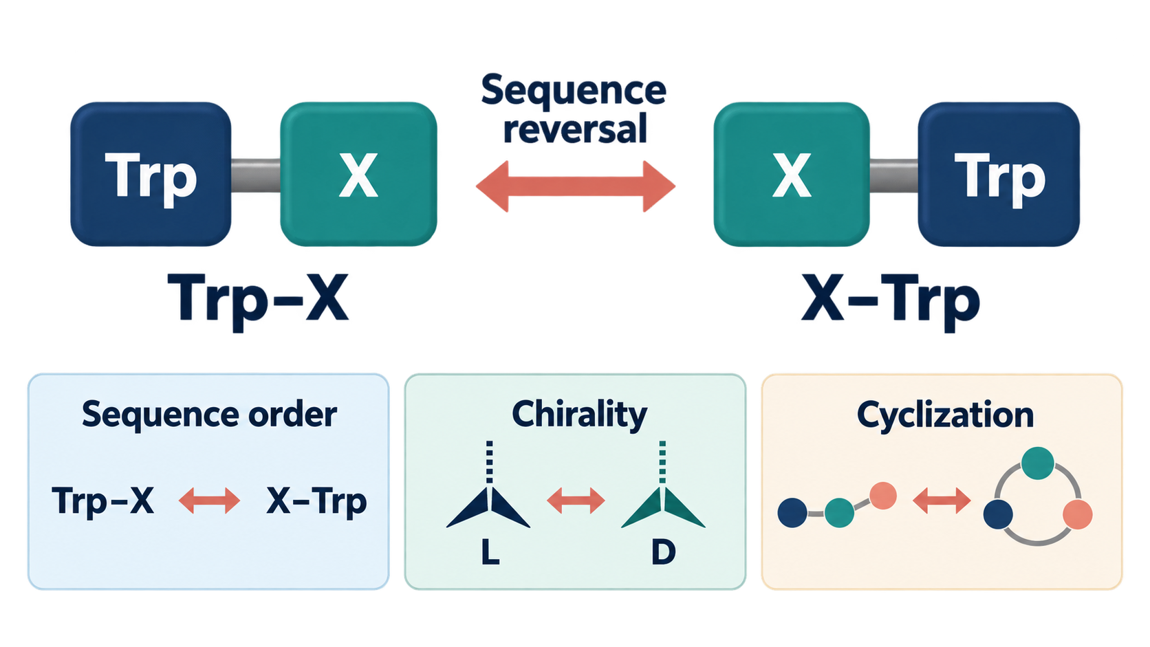
Sequence reversal changes residue order. It is not the same operation as changing L/D chirality or closing a chain into a ring. Conceptual illustration, not an exact molecular structure.
This is why the study supports a narrow conclusion: connectivity mattered in this library. It does not establish a universal rule that “Trp first” is better, or that reversing an unrelated peptide will predictably increase potency.
Sequence reversal is not chirality or cyclization
Residue order, mirror-image chirality and ring closure are different structural variables. Mixing them together makes peptide claims sound broader than the experiments actually are.
| Structural change | What changes | What it does not mean |
|---|---|---|
| Sequence reversal | Order/connectivity changes from Trp-X to X-Trp | The molecule has not automatically become a D-peptide or a ring |
| L/D chirality change | One or more stereocenters switch to a mirror-image configuration | The residue order can remain unchanged |
| Cyclization | A linear chain is closed through a backbone or side-chain connection | The sequence need not be reversed and the chirality need not change |
For more on the distinction, see our explainers on D-peptide mirror-image design and peptide cyclization.
What molecules did the researchers actually test?
These were novel, chemically modified tryptophan-derived dipeptides—not dietary tryptophan and not ordinary unmodified Trp-Pro. The library members carried N,N-dibenzyl substitutions and methyl ester groups; the reverse-series proline compound had a related but distinct substitution pattern.
That detail matters because the modifications can change lipophilicity, shape, steric constraints, cell exposure and interactions. Calling compound 4i simply “Trp-Pro” risks making readers think that any tryptophan-proline dipeptide, food source or supplement shares its activity. The paper’s full chemical name for 4i is methyl N,N-dibenzyl-L-tryptophyl-L-prolinate.
The work was a structure-activity relationship study: researchers varied the partner residue and its position, then compared assay results. It was not a nutrition study, supplement study or clinical drug trial.
What did the six-cell-line experiment measure?
The experiment measured a short-term change in cellular mass in cultured tumor-derived cells. A549, HeLa, MIA PaCa-2, SW1573, T-47D and WiDr are six laboratory cell lines derived from human tumors. They are not six groups of patients.
Cells were exposed to compounds for 48 hours. The researchers then used the sulforhodamine B colorimetric assay, which estimates cellular protein mass through dye binding. The study summarized effects as GI50: the tested concentration associated with 50% growth inhibition under the assay’s defined baseline and endpoint conditions. The broader NCI screening framework behind this measure is described by Monks and colleagues.
GI50 is not interchangeable with a direct count of dead cells. Less cellular mass after 48 hours could reflect slower proliferation, cell loss or a combination. Separate experiments would be needed to establish cell death, its mechanism and relevance in a living organism.
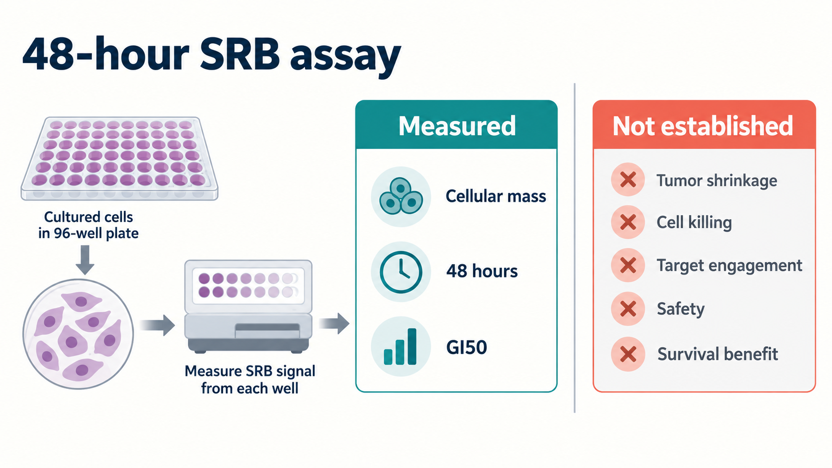
The SRB assay measured cellular mass after 48 hours and supported GI50 calculation. It did not establish tumor shrinkage, cell killing, target engagement, safety or survival benefit.
Measured versus not established
The paper provides phenotypic in-vitro growth-inhibition data; most questions needed for a treatment claim remain open.
| Measured or reported here | Not established by this study |
|---|---|
| 48-hour cellular-mass response in six tumor-derived cell lines | Tumor shrinkage in an animal or person |
| GI50 values for the tested library and reference drugs | Direct cell killing or a death mechanism |
| Means ± standard deviation from at least three independent experiments | Reproducibility across independent laboratories |
| Differences among paired Trp-X and X-Trp compounds | A universal rule that reversal improves potency |
| Phenotypic comparisons with cisplatin and 5-fluorouracil | Clinical superiority to chemotherapy |
| Activity in a tumor-cell panel | Normal-cell selectivity, pharmacokinetics, animal efficacy, human safety or survival benefit |
This is the practical boundary between an in-vitro signal and a medicine. Our guide to what preclinical actually means explains why cell data should remain at the cell-data level.
What does Table 1 show about compound 4i?
Compound 4i was the most consistently active member of this library, but its reported numbers are micromolar—not sub-micromolar. Table 1 gives the following mean GI50 values and standard deviations:
| Cell line | 4i GI50 (µM) |
|---|---|
| A549 | 1.36 ± 0.11 |
| HeLa | 1.59 ± 0.35 |
| MIA PaCa-2 | 2.00 ± 0.14 |
| SW1573 | 2.06 ± 0.47 |
| T-47D | 1.98 ± 0.24 |
| WiDr | 1.65 ± 0.38 |
The abstract, results discussion and conclusion describe 1.36–2.06 µM as “single-digit sub-micromolar.” Those words do not match the numbers: sub-micromolar ordinarily means below 1 µM. This article follows the table and reports the values as 1.36–2.06 µM. That is an internal wording-versus-number discrepancy, not evidence of misconduct.
Table 1 also prevents a blanket claim that 4i outperformed chemotherapy. In MIA PaCa-2 cells, 4i was 2.00 ± 0.14 µM, while cisplatin was 1.60 ± 0.27 µM. A lower GI50 indicates greater assay potency under those conditions, so 4i was not better in that comparison. In other lines, rankings varied.
Cisplatin and 5-fluorouracil were phenotypic assay references. They were not administered to patients in a randomized head-to-head trial. Cell-culture concentration comparisons cannot account for dosing, exposure, metabolism, toxicity, tumor biology or clinical outcomes.
Does Table 1 prove reversal usually improves potency?
No. The table shows a library-level tendency with important compound-specific exceptions, not a universal reversal rule. Many matched Trp-X compounds had lower GI50 values than their X-Trp counterparts, especially the proline pair 4i versus 8i. But other pairs moved differently.
For example, the phenylglycine X-Trp compound 8c had lower GI50 values than Trp-X compound 4c across the reported panel. The tyrosine pair also varied by cell line. The paper itself discusses a “rescue” of activity for selected residues after relocation.
That pattern is scientifically useful because it shows interactions among sequence position, residue identity and the library’s chemical modifications. It is also why “reversal improves potency” is too crude. The defensible summary is: reversal changed potency, and the direction depended on the compound and cell line.
Are the proposed mechanisms proven?
No direct molecular target was validated in the presented results. The paper discusses possible membrane interactions, transporter recognition, intracellular accumulation and conformational effects. These are hypotheses or interpretations, not demonstrated mechanisms for this library.
The results explicitly defer full mechanistic elucidation and target validation. The conclusion also says independently conducted follow-up work linked the leads to metabolic disruption and stress-induced autophagic flux, but those follow-up data are not presented in this paper. Until separately reported and evaluated, that statement should not be treated as evidence supplied by the current experiments.
What would need to happen next?
The next steps are target validation, selectivity testing and whole-organism pharmacology—not treatment use. Useful follow-up would include:
- confirming whether the compounds slow division, kill cells or do both;
- testing non-tumor human cell models to estimate selectivity;
- identifying and validating molecular targets or transport pathways;
- measuring stability, exposure, distribution and clearance;
- testing efficacy and toxicity in appropriate animal models;
- reproducing key findings independently; and
- only after adequate preclinical development, evaluating safety and benefit in phased human trials.
None of those stages is guaranteed to succeed. Cell-active compounds frequently fail because they cannot reach useful exposure, lack selectivity, behave differently in organisms or create unacceptable toxicity.
Frequently asked questions
Does reversing a peptide always change its activity?
It can, but the size and direction of the change are molecule- and assay-specific. This study cannot supply a rule for all peptides.
Is sequence reversal the same as making a D-peptide?
No. Sequence reversal changes residue order. A D-peptide changes stereochemistry. Researchers can alter either variable without altering the other.
Did the study test Trp-Pro supplements?
No. Compound 4i was a chemically modified peptidomimetic with N,N-dibenzyl and methyl ester groups, not ordinary dietary tryptophan, a supplement or unmodified Trp-Pro.
Do six human tumor cell lines mean six human trials?
No. They are cultured cell models derived from human tumors. No patients were treated.
Does a low GI50 prove cancer cells were killed?
No. GI50 describes growth inhibition under defined assay conditions. The SRB endpoint measured cellular mass, not a specific cell-death mechanism.
Was compound 4i sub-micromolar?
Not according to Table 1. Its reported GI50 range was 1.36–2.06 µM. The paper’s “sub-micromolar” wording conflicts with those values.
Bottom line
The Díaz study is a useful demonstration that connectivity matters in medicinal chemistry. Reordering two residue positions changed both chemical behavior and cell-growth effects across a modified tryptophan-derived dipeptide library.
Its strongest lesson is evidence literacy, not cancer-treatment advice. The work used a 48-hour cellular-mass assay in six tumor-derived cell lines. It did not establish tumor shrinkage, clinical superiority, a validated target, normal-cell selectivity, pharmacokinetics, animal efficacy, safety or patient benefit.
This article is for general education and evidence literacy. It is not medical advice and does not provide dosing, purchasing, sourcing or treatment instructions.
Sources
- J. Benamer Díaz, Adam N. Khan, Aday González-Bakker, and José M. Padrón. “Sequence Inversion Dictates the Antiproliferative Activity of Bidirectional Tryptophan-Containing Dipeptide Libraries.” Journal of Peptide Science. First published September 16, 2026. https://doi.org/10.1002/psc.70128
- Vichai V, Kirtikara K. “Sulforhodamine B Colorimetric Assay for Cytotoxicity Screening.” Nature Protocols. 2006. https://doi.org/10.1038/nprot.2006.179
- Monks A, Scudiero D, Skehan P, et al. “Feasibility of a High-Flux Anticancer Drug Screen Using a Diverse Panel of Cultured Human Tumor Cell Lines.” Journal of the National Cancer Institute. 1991. https://doi.org/10.1093/jnci/83.11.757