Does Closing a Peptide Into a Ring Make It a Better Drug? What a New Crosslinking Study Shows
Quick answer
Closing a peptide into a ring can improve particular laboratory properties, but it does not automatically make a better drug. A peptide might resist breakdown yet bind less effectively, or enter a cultured cell without reaching a useful intracellular target. Stability, binding, cell entry and clinical benefit need separate evidence.
That distinction is central to Liu and colleagues’ Nature Communications study, published September 2, 2026. The researchers linked amino-acid side chains to create cyclic peptides, then tested different peptide families in different laboratory assays. Some comparisons improved; others did not.
This is a chemistry and in-vitro study, meaning its biological experiments used laboratory systems and cultured cells. It is not a human cancer-treatment trial. Its lasting lesson is how to read evidence about cyclic peptides—not which experimental compound to use.
In this article
- How side-chain crosslinking makes a ring
- Stability, binding and the evidence ladder
- What improved and what did not
- Uptake, apoptosis and normal-cell findings
- Study limitations
- What evidence is needed next
How does side-chain crosslinking make a peptide ring?
A bridge between two side chains creates a loop and reduces the shapes a peptide can adopt. Think of a flexible string with two short branches attached at different points. Connecting those branches closes a loop while leaving the string’s ends free.
A peptide is a chain of amino acids. Each amino acid has a side chain extending from the backbone. In the central approach studied here, two lysine side chains become connected through a urea- or thiourea-containing bridge. The researchers also made versions with a bridge containing two thiourea groups. These names describe the chemical connection, not a level of clinical effectiveness.
This is different from simply joining the two ends of a peptide. A side-chain crosslink can enclose part of the backbone within a ring while other parts remain outside it. The result is constrained, not necessarily locked into one perfectly rigid shape.
Why might that help? A constrained peptide may spend more time in a shape suited to a binding partner, or become harder for a breakdown enzyme to recognize. But it might also become less able to adopt the needed shape. The bridge itself changes chemical interactions as well as geometry. Consequently, “cyclic versus linear” is not a test of shape alone.
Sequence order is another distinct design variable: reversing modified dipeptides from Trp-X to X-Trp changed 48-hour cell-growth readouts, but did not establish a cancer treatment.
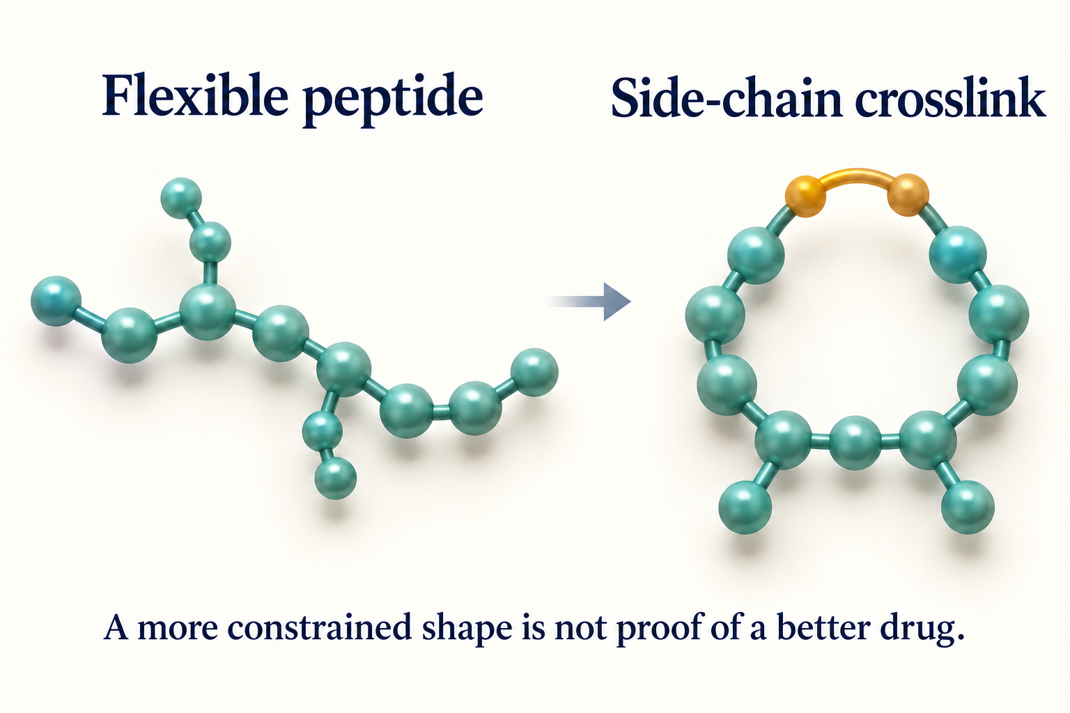
A side-chain bridge restricts shape without joining the chain ends. Conceptual illustration, not a molecular structure or study result.
Cyclic peptides: stability, binding and the evidence ladder
Each assay answers a different question; success in one does not establish success in the others. The evidence ladder here runs from chemical characterization and laboratory measurements toward whole-body and human outcomes. The study does not reach those last stages.
Importantly, its laboratory results are not consecutive milestones achieved by one compound. They come from several families:
- Stability: P24 versus 24a and 24b. These HHC-36-related peptides were compared under oxidative, alkaline and trypsin challenges. The measurements ask how much peptide remains under those conditions—not how long a medicine lasts in a person.
- Integrin binding: P26 versus 26b. These RGD-containing peptides were tested in a competitive assay involving integrin αvβ6, a protein that interacts with extracellular binding partners. This measures interference with a particular protein interaction.
- Cell-growth effects: P23 and P24 families. Anoplin-analogue derivatives 23a–23c and HHC-36 derivatives 24a–24b were compared with their linear parents in cancer-derived cell lines.
- Fluorescent uptake: P25 versus 25a–25c. This separately labeled peptide family was examined for cell-associated and intracellular fluorescence. It is not interchangeable with the compounds in the growth experiments.
- Docking: proposed protein interactions. Computer models explored possible binding poses, including 23c with EGFR. A modeled pose is a hypothesis, not observed target engagement.
Clinical benefit would require evidence that a treatment improves meaningful outcomes in people, with an acceptable balance of benefit and harm. None of these laboratory readouts supplies that answer. Our guide to what preclinical actually means explains that boundary in more detail.
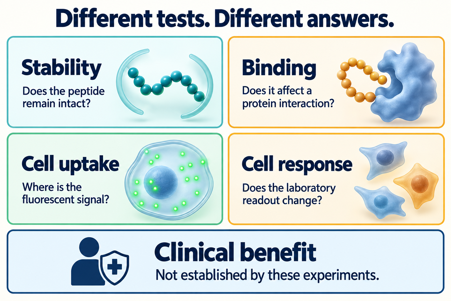
These measurements answer different questions and used different peptide families. They are not a single compound’s progression toward clinical success.
What improved, and what did not?
The results support selected improvements—not a rule that every cyclic analogue is better. Both the peptide family and the cell line change the conclusion.
Stability improved in the tested HHC-36 comparisons
The cyclic HHC-36 derivatives generally retained more material than linear P24 under the reported challenges. The supplementary information, Figures S39–S45, shows the associated chromatographic evidence.
The source workbook also prevents an overly absolute description. Its oxidative-stability series decreases for 24b, even though it remains above P24. “More resistant under this challenge” is a better summary than “does not degrade.” Resistance to one enzyme or chemical environment is not resistance to every breakdown process in a body.
The RGD-containing cyclic peptide improved competitive inhibition
The paper reports a lower IC50 for 26b than for linear P26 in the integrin assay, approaching the cyclic RGD positive control. IC50 means the concentration associated with half-maximal inhibition in that assay. A lower value indicates greater inhibitory potency under the assay conditions; it is not automatically a direct binding constant.
This was a competitive ELISA involving integrin αvβ6 and its binding partner LAP. The experiment included a cyclic RGD reference compound, background wells without integrin, and total-binding wells without competing RGD derivatives. Those controls help interpret the signal. They do not turn it into evidence of tumour shrinkage or human benefit.
Cell-growth results included clear reversals
Table 1 gives the most useful corrective to the paper’s broad language:
- 23c improved over P23 in A549-Luc and Caco-2 cells, but not in A498 cells. In A498, its reported IC50 was about 2.8 times higher than P23’s, indicating weaker antiproliferative activity in that comparison.
- 23a and 23b performed worse than P23 in A549-Luc cells. All three tested cyclic P23 derivatives also had higher reported IC50 values than P23 in A498.
- 24b had lower reported IC50 values than P24 across all three cell lines. The improvements for 24a were smaller or absent, depending on the line.
The article’s narrative says 23c was more potent than P23 across all three lines. Its own Table 1 does not support that statement. This explainer follows the table’s comparison rather than repeating the broader claim.
These are assay-specific rankings, not treatment rankings. A549-Luc, Caco-2 and A498 are cancer-derived cell models; an effect in any of them is not proof of benefit for a person with that cancer.
What do uptake, apoptosis and normal-cell tests show?
They add different kinds of information, but none establishes successful drug delivery or systemic safety. Keeping their endpoints separate avoids turning several limited findings into one oversized conclusion.
Fluorescence supports uptake, not oral absorption
The labeled cyclic P25 derivatives produced stronger fluorescence than the labeled linear parent in A549-Luc experiments, with 25b showing the strongest reported enhancement. Microscopy and flow cytometry support an uptake-related difference under the tested conditions.
However, a fluorescent label is not a measurement of intact, active peptide at its intended intracellular target. Cell-associated signal, intracellular location, peptide integrity and biological action are distinct questions. The label itself is also part of the tested molecule.
The supplement includes a separate viability assay for this labeled family, with reduced viability for some analogues. That matters when interpreting uptake: a brighter signal cannot simply be treated as evidence of harmless membrane passage. The viability and uptake readouts also cover different exposure windows.
Nothing here measures absorption after swallowing, passage into blood, or oral bioavailability. For a separate agent-specific discussion, see LUNA18/paluratide and oral cyclic-peptide evidence. Evidence about that molecule does not transfer to these compounds.
Apoptosis markers are not the same as reduced cell growth
The main cell-growth screen used CCK-8, a metabolic viability readout. A lower signal can reflect fewer viable cells or changes in their metabolic activity; it does not by itself identify a cell-death mechanism.
A separate experiment examined 23c using Annexin V and propidium iodide in A549-Luc and Caco-2 cells. The authors reported increased apoptosis-associated populations relative to untreated controls. The methods describe blank and single-stain controls, which help interpret the fluorescence measurements.
That adds evidence consistent with cell death in those models. It does not demonstrate that EGFR caused the effect or that a tumour would respond in a living organism.
One normal-cell result is not a safety profile
The authors report that selected compounds 23c and 24b maintained more than 80% MCF-10A viability at their antitumour IC50 concentrations. MCF-10A is a non-tumour breast epithelial cell model. That is a limited comparison with the cancer-cell findings, not evidence that the compounds spare every healthy tissue.
There is an additional qualification: the source workbook contains a discrete concentration series, not a separate measurement at every fitted cancer-cell IC50. The statement is therefore best read as the authors’ interpretation of those curves. The same normal-cell data show substantial loss of viability for 23c at higher tested concentrations.
A single cell line cannot establish effects on organs, immunity, distribution, metabolism or longer-term exposure. “Some separation in these cell assays” is defensible; “systemically safe” is not.
What are the study’s main limitations?
The study supplies useful laboratory comparisons, but small datasets, reporting inconsistencies and missing mechanistic evidence limit their reach. Reading the supporting files makes those boundaries clearer.
The reporting summary states that no sample-size calculation was performed. It describes independent biological experiments and lists randomization and blinding as not applicable. Cell lines were used without further authentication in the authors’ laboratory; the authors report negative mycoplasma tests.
Replicate counts are not identical everywhere. Integrin results are reported with three biological replicates, and growth-assay captions generally state at least three. The source-data workbook labels some growth datasets as four or three-to-four replicates, despite the reporting summary’s general description of three. These are small laboratory replicate sets, not groups of patients.
The workbook includes normalized viability and inhibition values. Some viability series are not smoothly concentration-dependent, and some normalized values exceed 100%. That does not mean cells are “more than completely alive”; it means the readout is relative to a control. It also argues against treating fitted IC50 values as perfectly precise rankings. The stability sheets show single value series rather than separate replicate columns, and the workbook does not contain raw flow-cytometry event data for uptake or apoptosis.
Finally, docking does not confirm EGFR target engagement. EGFR was selected as a candidate through computational analysis, then modeled with P23 and 23c. This proposes a possible explanation. It does not experimentally demonstrate binding inside cells, inhibition of EGFR, or dependence of the cell-growth effect on that target. The distinction parallels the broader gap between peptide binding and biological effect. A separate TREM2 cyclic-peptide study likewise shows why measured binding plus cell-model changes still leaves delivery and organism-level efficacy unresolved.
What evidence would be needed next?
A stronger drug-development claim would need reproducible, connected evidence for the same candidate—not a collection of wins from different peptide families. The missing knowledge includes whether the comparisons reproduce independently, whether the intact compound reaches the relevant cellular compartment, and whether its proposed target actually accounts for the observed effect.
Whole-body exposure, distribution, breakdown products and toxicity would also need characterization before laboratory stability could be translated into a pharmacological claim. Any eventual claim of clinical benefit would require appropriate human evidence. This study does not establish that these steps will succeed or provide a timeline for them.
The practical reading rule is simple: ask which peptide, which measurement, which comparator, and which model? Ring closure can be a useful design tool. The evidence—not the ring—determines whether it produces a better medicine.
This article is educational and does not provide treatment, preparation or self-experimentation advice.
Sources
The primary article, its supplementary assay details, reporting summary and source workbook were checked for this explainer.
- Liu et al., “Macrocyclization of native peptides through (Thio)urea crosslinking of two amines,” Nature Communications, September 2, 2026. DOI: 10.1038/s41467-026-77301-w. Peer-reviewed Article in Press.
- Full article PDF, especially Table 1 and Figures 7–9.
- Supplementary information, especially sections H–P and Figures S45–S60.
- Reporting summary, study design, cell-line reporting and flow cytometry.
- Source data workbook, including the Table 1 and Figures S45–S51 sheets.