Can Ancient Lactoferrin Peptides Kill Drug-Resistant Bacteria? What the Resurrection Study Actually Shows
Ancient antimicrobial peptides reconstructed from mammalian lactoferrin damaged bacterial membranes and, in later reconstructed forms, killed several laboratory strains more effectively. That is a useful evolutionary finding and a possible starting point for drug discovery. It is not evidence that an ancient peptide can treat an infection in a person.
The distinction matters because the August 25, 2026 PLOS Biology study was a sequence-reconstruction and laboratory-assay paper. Researchers inferred extinct protein sequences, synthesized 25-amino-acid peptide segments, and tested them against bacteria in controlled media. They did not test an antibiotic in animals or humans.
Quick answer: The study shows that membrane-disrupting activity appeared early in the evolution of lactoferricin and that stronger bactericidal activity emerged in later ancestral and living mammalian variants. It does not establish dosing, clinical safety, effectiveness in an infected body, or activity against patient-derived multidrug-resistant infections.
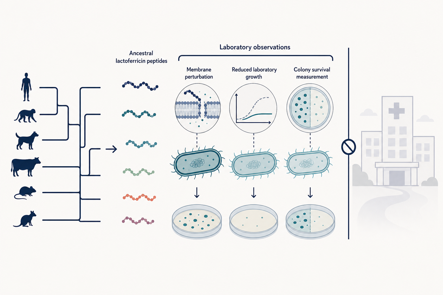
The study connects reconstructed sequences to laboratory observations. A substantial evidence gap remains between those experiments and a validated treatment.
What did the lactoferricin resurrection study find?
The central result was an evolutionary progression—not the discovery of a ready-made antibiotic.
The researchers reconstructed lactoferrin ancestors around key branches in mammalian evolution. They then synthesized the conserved 25-amino-acid region corresponding to lactoferricin and compared those peptides with human and bovine lactoferricin and with matching transferrin segments.
The earliest reconstructed lactoferricin, AncLFcin1, could permeabilize Pseudomonas aeruginosa membranes and alter membrane potential, yet its effect on growth and survival was relatively weak or recoverable. AncLFcin2 and AncLFcin3 generally showed stronger growth inhibition and bactericidal activity. Living human lactoferricin was active in several assays, while bovine lactoferricin was the most consistently potent variant tested.
This was not a smooth march toward greater potency. Some reconstructed ancestors were more active than human lactoferricin against Staphylococcus aureus. Activity depended on both the peptide and the bacterial species.
What is ancestral sequence reconstruction?
Ancestral sequence reconstruction, or ASR, is a statistical estimate of an extinct protein sequence—not a recovered ancient molecule.
The team used the Topiary pipeline and 376 vertebrate transferrin-family sequences to build a gene tree and infer the most likely amino acid at each ancestral position. The approach combined a multiple-sequence alignment, a maximum-likelihood gene tree, reconciliation with the species tree, and posterior probabilities for individual residues.
Because some positions were uncertain, the researchers also created “Altall” versions. At ambiguously reconstructed sites, defined using Topiary's default posterior-probability cutoff of 0.25, the alternate residue replaced the most likely one. These alternate peptides produced broadly similar antimicrobial patterns against P. aeruginosa PAO1 and S. aureus JE2. One alternate ancestral transferrin segment showed weak activity against S. aureus only at the highest tested concentrations.
That robustness check is useful, but it does not make the inferred sequences certain. ASR answers, “What sequence is most compatible with this evolutionary model and these modern sequences?” It does not provide a literal sample from an extinct mammal.
Lactoferrin and lactoferricin are not the same thing
Lactoferrin is a large iron-binding protein; lactoferricin is a short antimicrobial domain that can be released from its N-terminal region by proteolytic cleavage.
Lactoferrin occurs in milk, tears, saliva, neutrophil granules, and other mammalian secretions. The intact protein has functions that include binding iron. Lactoferricin is one embedded segment within it. Host proteases such as pepsin, trypsin, and chymotrypsin can liberate lactoferrin-derived peptides, but the conditions and resulting fragments vary.
The experiments did not administer reconstructed full-length lactoferrin to infected organisms. They used commercially synthesized 25-amino-acid peptide segments corresponding to residues 17–42 of mature human lactoferrin. Claims about these short peptides should not be silently transferred to intact lactoferrin, and claims about intact lactoferrin should not be assumed to describe isolated lactoferricin.
That separation is one example of how to evaluate peptide claims without letting one experiment carry more weight than it can support.
Which bacteria, concentrations, and assays were tested?
The broad screen covered six laboratory strains, followed by deeper experiments with PAO1 and JE2 and additional screens of several staphylococci.
The dose-response growth experiments tested:
- Pseudomonas aeruginosa PAO1 and PA14
- Escherichia coli ATCC 25922
- Staphylococcus aureus JE2 and MN8
- Streptococcus agalactiae MNZ933
Peptides were screened mainly from 50 to 800 micrograms per milliliter, with the paper describing the broader experimental range as 0.1–1 milligram per milliliter. Growth was monitored by optical density for 24 hours in low-nutrient 5% tryptic soy broth. The researchers summarized growth using area under the curve.
For deeper survival experiments, PAO1 received 100 micrograms per milliliter and JE2 received 800 micrograms per milliliter. Viable bacteria were measured by colony-forming units over time. The authors also checked selected trends in 0.5% milk and Opti-MEM.
Most main figures report three biological replicates. Where significance was assessed, the paper used one-way ANOVA and displayed thresholds from P ≤ 0.05 to P ≤ 0.0001. The microscopy cell-size analysis measured 100 cells. These choices support a controlled laboratory comparison; they do not substitute for replication across clinical isolates, infection models, or trials.
Evidence check: Optical density indicates how cloudy a culture becomes, not necessarily whether every bacterium is alive. Colony counts more directly assess survival. Propidium iodide, membrane-potential dye, Nile Red microscopy, and cell-size measurements address membrane effects. Each assay answers a different question.
Did the early peptides kill bacteria or only disturb membranes?
The earliest reconstructed domain clearly perturbed membranes, but its killing was weaker and sometimes followed by bacterial recovery.
All tested ancestral and living lactoferricin peptides allowed propidium iodide into PAO1 cells within 30 minutes and changed membrane polarization. Matching transferrin-derived peptides did not. Microscopy showed membrane puncta and smaller cells after ancestral peptide exposure, with more severe deformation after human and especially bovine lactoferricin.
Survival assays supplied the more important distinction. AncLFcin1 caused an early drop in PAO1 viability followed by partial recovery. AncLFcin2 produced roughly a 1,000-fold decrease after 24 hours. Human lactoferricin produced a larger late reduction, while bovine lactoferricin left no detectable PAO1 colonies within two hours under that assay condition.
Against JE2, AncLFcin2 reduced viability by about 100,000-fold by the end of treatment. Human lactoferricin initially reduced viability before the culture resumed growth. Bovine lactoferricin again eliminated detectable colonies within two hours.
“No detectable colonies” means below the detection limit of that experiment. It does not mean sterilization in every environment or cure of an infection.
What did the Q5R experiment actually show?
Replacing glutamine with arginine at position 5 increased the potency of human lactoferricin in these assays, particularly against S. aureus. It did not validate an antibiotic.
Humans and many monkeys carry glutamine at lactoferricin position 5, whereas other great apes carry arginine. The researchers synthesized human lactoferricin with the Q5R substitution and compared it with ordinary human lactoferricin. They also tested a K12R variant at another rapidly evolving site.
Q5R significantly improved activity against S. aureus JE2 and suppressed growth across additional staphylococcal isolates. The raw supporting workbook identifies these as S. lugdunensis, S. hominis, S. caprae, and S. haemolyticus. The article text and figure caption contain inconsistent species labels in this part of the paper, so the raw-data sheet names are the clearest record of what was analyzed.
K12R behaved similarly to ordinary human lactoferricin against PAO1 and JE2, although the authors noted modest growth reductions in some broader screens.
Q5R adds a positively charged arginine next to a conserved aromatic residue, a plausible way to strengthen interaction with negatively charged bacterial surfaces and insertion into membranes. But a one-residue improvement in a culture assay does not establish stability in blood or tissues, distribution, immune effects, resistance risk, formulation, animal efficacy, or human safety.
Did the researchers test safety?
They performed one narrow hemolysis screen, not a therapeutic safety program.
Bovine red blood cells were exposed to each peptide at 1 milligram per milliliter for one hour. Hemolysis was compared with a 1% Triton X-100 positive control, and no significant hemolysis was reported for the tested peptides across three biological replicates.
This is encouraging only within its limited scope. A red-blood-cell assay can detect one form of membrane toxicity. It cannot rule out toxicity to other cell types, immune reactions, organ injury, altered microbiomes, off-target effects, or harm after exposure in a living animal or person.
Does this study show ancient antimicrobial peptides can treat resistant infections?
No. The work identifies experimentally interesting peptide variants, but therapeutic evidence begins several steps later.
This is the same boundary explained in our guide to what preclinical evidence actually means. It also distinguishes this work from other experimental peptide strategies aimed at antibiotic resistance, which face their own delivery and validation problems.
The paper did not report:
- animal infection studies
- human pharmacokinetics or safety
- clinical trials
- comparison with standard antibiotics
- testing across a defined panel of multidrug-resistant clinical isolates
- resistance-evolution experiments
- formulation or delivery performance
The tested species include pathogens that can be drug resistant, but a species name is not a resistance phenotype. PAO1, PA14, JE2, MN8, ATCC 25922, and MNZ933 should not be described collectively as proven multidrug-resistant clinical isolates without susceptibility data from this study.
The authors also emphasized that most work used low-nutrient laboratory medium that does not fully reproduce a host environment. pH, salts, proteins, proteases, and other chemical conditions can substantially change antimicrobial-peptide activity. The general patterns persisted in two alternative media, but that remains in vitro evidence. A cobra-inspired antimicrobial-peptide screen provides a complementary example of why computational promise, culture activity, and therapeutic viability must remain separate claims.
The bottom line
The 2026 study offers a strong evolutionary lesson: a cryptic antimicrobial domain inside lactoferrin appears to have acquired membrane-disrupting properties early, with later sequence changes increasing bactericidal activity in ways that depended on the target bacterium. Alternate ancestral reconstructions supported the main pattern, and Q5R showed that one naturally varying site can materially change potency.
It also offers an evidence-literacy lesson. Sequence inference is not a fossil. Membrane damage is not the same as bacterial death. Killing laboratory strains is not the same as treating an infection. A hemolysis check is not comprehensive safety evidence. Ancient antimicrobial peptides may be useful leads for future research, but this paper did not turn one into a medicine.
Frequently asked questions
Was full-length ancient lactoferrin tested against bacteria?
No. Full-length reconstructed proteins were modeled structurally, but the antimicrobial experiments used synthesized 25-amino-acid lactoferricin-region peptides.
Were the bacteria definitely drug resistant?
The study used recognized laboratory strains from pathogenic species, but it did not establish that the full panel represented multidrug-resistant clinical infections. The results should be described as activity against tested laboratory strains.
Does Q5R make human lactoferricin an antibiotic?
No. Q5R increased potency in specified in-vitro assays, especially against S. aureus. A validated antibiotic requires much broader preclinical and clinical evidence.
Did the peptides damage red blood cells?
The researchers reported no significant hemolysis after a one-hour bovine red-blood-cell assay at 1 milligram per milliliter. That is a narrow laboratory safety check, not proof of safety in animals or humans.
Sources
- Sil T, Kowalski CH, Scamfer S, Copeland N, Barber MF. “Retracing the origin and evolution of a cryptic antimicrobial peptide within mammalian lactoferrin.” PLOS Biology. Published August 25, 2026.
- The paper's open supporting information: S1–S8 figures and S1–S3 data, including raw assay tables, alternate ancestral reconstructions, the reconstruction tree, and reconstructed sequences.
This article is educational and does not provide infection-treatment or medical advice.