Why ‘Not Found in Healthy Tissue’ Does Not Prove a Peptide Cancer Target Is Safe
A peptide cancer target is not proven safe just because researchers cannot find it in a healthy-tissue database. A database can only report what its samples, HLA alleles, cell types, biological states, instruments, and search methods were capable of detecting. “Not detected” is therefore a qualified observation—not proof that the peptide-HLA target is absent from every normal human tissue.
That is the central evidence-literacy lesson of a September 1, 2026 paper in the Journal for ImmunoTherapy of Cancer (JITC), “Normal-immunopeptidome denominator: a safety atlas for immunotherapies targeting non-canonical and aberrantly expressed tumor antigens”. The author proposes a disqualification-first framework for judging peptide-HLA targets before therapeutic nomination.
What this paper is: an open-access, externally peer-reviewed framework/commentary published as a journal article and indexed by PubMed as a review. It proposes how evidence should be assembled and interpreted.
What it is not: a clinical trial, a treatment result, a completed or clinically validated safety atlas, or proof that applying the framework makes a target safe.
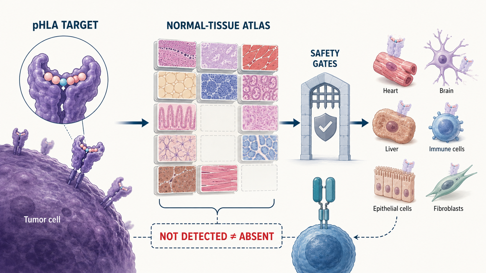 A normal-tissue database can help disqualify a target, but gaps in sampling and detection mean that “not detected” is not the same as “absent.”
Quick facts
- The target is a surface complex. HLA molecules display short peptides on cells; a therapy may recognize a particular peptide-HLA, or pHLA, combination.
- Origin and distribution are different questions. “Non-canonical” describes where a peptide came from at the molecular level. It does not say where the resulting pHLA appears in the body.
- RNA and protein are indirect clues. They do not establish which pHLA complexes are naturally presented on a cell surface.
- A normal-immunopeptidome denominator is a filter, not a safety certificate. It compares candidates with measured pHLA presentation across benign tissues and states.
- The intended therapy changes the risk calculation. A vaccine, a persistent TCR-T cell, and a TCR-based bispecific do not have the same sensitivity or therapeutic index.
What is a peptide-HLA target?
A peptide-HLA target is a short peptide displayed in the binding groove of a human leukocyte antigen molecule on the surface of a cell. T cells survey these pHLA complexes. Cancer immunotherapies can try to direct that recognition toward tumor cells.
The peptide is only one half of the address. The HLA molecule is the other. The same peptide paired with a different HLA is not necessarily the same therapeutic target, and a target relevant to one HLA allele may not apply to patients with another.
This is why target evaluation has to focus on naturally presented pHLA complexes, not simply on whether a gene is transcribed or a protein is abundant. Between RNA and surface presentation sit many biological steps: translation, protein turnover, proteolytic cleavage, intracellular transport, HLA binding, peptide editing, and changes in cellular state.
The paper gives two useful counterexamples:
- A transcript that looks absent at baseline could still generate a pHLA under a particular stress or inflammatory state.
- An abundant protein might never produce the exact surface pHLA a therapeutic receptor recognizes.
For a broader guide to separating direct measurements from biological inference, see how to evaluate peptide claims online.
Canonical versus non-canonical does not mean safe versus unsafe
Molecular origin and normal-tissue distribution are separate axes. Mixing them up is the basic category error behind many “novel target” headlines.
| Question | Main categories | What the classification tells you |
|---|---|---|
| Where did the peptide sequence come from? | Canonical or non-canonical | Its molecular source or translation context |
| Where is the pHLA presented? | Tumor-specific, tumor-associated, or lineage-specific | Its distribution and selectivity across tumor and normal tissues |
A canonical peptide generally comes from a conventionally annotated protein-coding sequence. A non-canonical peptide can arise from less conventional sources, such as non-coding regions, alternative reading frames, splice products, or other atypical translation events.
The distribution labels answer a different question:
- Tumor-specific antigen (TSA): presented by tumor cells and not shown to be presented by normal tissues in the relevant context.
- Tumor-associated antigen (TAA): enriched or altered in tumors but also present in at least some normal context.
- Lineage-specific antigen: tied to a cell lineage that may include both malignant and non-malignant cells.
The paper’s point is explicit: a non-canonical pHLA may be shared with benign tissue, while a canonical pHLA may show tumor-specific presentation. “Non-canonical” can make a candidate scientifically interesting. It cannot make the candidate safe by definition.
Why a missing healthy-tissue database hit is weak negative evidence
A negative database search only excludes what the database was equipped to see. For peptide cancer target safety, immunopeptidome coverage matters at least as much as the search result.
Existing resources cover different parts of the problem. The paper notes that the HLA Ligand Atlas reported paired HLA class I and II data from 227 benign-tissue samples. PCI-DB integrates millions of class I and II peptides from malignant and benign primary tissues across more than 40 tissue types and cancer entities. IEAtlas helps annotate epitopes from non-coding regions, while Ligand.MHC Atlas supports evidence about natural tumor presentation and recurrence.
Those are useful layers, but they are not interchangeable:
- A source-oriented atlas can support where a peptide may have originated without proving presentation in the intended tumor.
- A tumor-focused atlas can support recurrence across samples without proving safety in normal tissue.
- A benign-tissue atlas can reveal normal presentation without ruling out receptor cross-reactivity with a different peptide.
Coverage is uneven across organs, cell types, HLA alleles, ancestries, and physiological states. Methods also differ in tissue handling, HLA purification, mass-spectrometry depth, acquisition mode, search-space construction, and false-discovery-rate control.
Normal baseline samples create another blind spot. A pHLA may be inducible during inflammation, interferon signaling, infection, hypoxia, aging, tissue repair, cellular senescence, radiation exposure, or drug treatment. A database built mostly from quiet baseline tissue cannot automatically answer what happens when biology stops being quiet—which it does with impressive regularity.
What is the proposed normal-immunopeptidome denominator?
The proposed denominator is a context-aware reference of pHLA ligands measured across benign human tissues, cells, HLA alleles, and biological states. Candidate tumor targets are the numerator. The normal-tissue presentation landscape is the denominator against which those candidates must be tested.
The purpose is primarily to disqualify unsafe-looking candidates early. If the same target pHLA is convincingly presented by a vulnerable normal tissue, that is highly relevant evidence against advancing it.
But the author argues that the denominator should produce a graded, auditable evidence dossier—not a simple blacklist. Each result should retain details such as:
- donor and tissue source;
- HLA assignment;
- sample-processing and acquisition method;
- search-space definition;
- spectrum-level evidence and false-discovery-rate controls;
- quantitative context, where available;
- what tissues, alleles, ancestries, cell states, and inducible conditions were not tested.
That provenance prevents broad claims from being built on narrow sampling. It also allows a target assessment to change when deeper or more representative benign-tissue data arrive.
The five core gates for target nomination
The framework requires five core gates before a pHLA target is nominated for therapeutic development. Passing one gate does not substitute for the others.
1. Is the peptide identification analytically credible?
The sequence, HLA assignment, and proposed molecular source need defensible analytical support. Search spaces and false-discovery-rate controls should be transparent. This matters especially for non-canonical searches because the candidate universe becomes much larger and one peptide can map to several transcripts, reading frames, splice isoforms, or homologous loci.
An exciting sequence with ambiguous source assignment or weak spectral evidence is not a stable foundation for therapy.
2. Is the pHLA naturally presented on tumor cells?
The target should be demonstrated on the intended tumor as a pHLA, using mass spectrometry or a validated orthogonal assay. RNA expression and total protein abundance can provide context, but they are not direct pHLA evidence.
Independent confirmation becomes more important when the initial identification is uncertain or the consequences of a mistake are high.
3. How much target is present, in how many tumors, and in which patients?
Where the data permit, investigators should assess pHLA abundance, distribution, and heterogeneity. A target found at useful density in one sampled region may be scarce elsewhere in the same tumor or absent from another patient’s tumor.
The dossier should also state whether the target is:
- patient-specific, potentially narrowing relevance but allowing personalization; or
- shared, potentially increasing coverage while raising the importance of broad normal-tissue scrutiny.
Heterogeneity connects directly to immune escape. Tumor cells that do not present the target—or that lose antigen-processing or HLA-presentation machinery—may evade a target-specific immune response.
4. What does the normal-immunopeptidome denominator show?
Investigators should search relevant benign pHLA datasets and document the coverage gaps. The conclusion should not be “safe because absent.” It should look more like: not detected under these methods, in these tissues and states, for these HLA alleles, at this depth, with these remaining uncertainties.
That wording is less cinematic. It is also what the evidence actually supports.
5. Is recognition selective enough for the intended modality?
The final core gate requires functional evidence that the therapy recognizes tumor cells while sparing appropriate normal cells, followed by a modality-specific therapeutic-index decision.
The same target evidence can support different decisions for different therapeutic formats:
| Modality | Why target evidence may be interpreted differently |
|---|---|
| Vaccine | Depends on endogenous immune priming and may not reach the sensitivity of an engineered high-affinity receptor |
| TCR-T cell | Cells can persist and expand, so weak unintended recognition can carry serious consequences |
| TCR-based bispecific | Can recruit many effector cells toward even low-density pHLA, increasing both potential activity and off-target concern |
Too little tumor pHLA may limit activity. Too much sensitivity to low-level or unintended normal-cell pHLA may narrow safety. The relevant question is not whether a target exists; it is whether the intended modality has a workable window between tumor recognition and normal-tissue harm.
The same endpoint discipline applies after treatment begins: our review of personalized pancreatic cancer vaccines explains why detecting an immune response does not by itself establish longer survival or clinical benefit.
What additional tests become necessary when risk is higher?
The framework escalates testing when the biology or therapeutic modality creates extra uncertainty. These are not decorative “nice to have” experiments when the corresponding risk applies.
If normal tissues might increase target presentation, the paper calls for prospective inducibility testing under relevant conditions such as interferon signaling, infection, epigenetic therapy, radiation, or tissue injury.
For TCR-T cells, affinity-enhanced TCRs, and TCR-based bispecifics, receptor-level safety evaluation should include:
- peptide-library screening;
- scans for homologous peptides;
- broader primary normal-cell panels;
- HLA alloreactivity testing, which checks whether the receptor reacts to other HLA contexts;
- investigation of molecular mimics that may differ from the intended peptide but still fit the receptor.
The danger is not limited to the exact target appearing in normal tissue. A receptor may cross-react with another pHLA. The paper points to earlier MAGE-A3 programs in which unexpected normal-tissue or cross-reactive recognition was associated with severe cardiac and neurological toxicity. Those cases are historical warnings, not results of the 2026 framework paper.
Which measurements could reduce the remaining uncertainty?
More precise measurement can narrow uncertainty, but it cannot turn incomplete evidence into certainty. The paper recommends several enhancements and says they should become required when material uncertainty remains:
- absolute quantification of pHLA density;
- high-resolution single-cell or spatial profiling;
- longitudinal sampling;
- organ-specific primary-cell or organoid models.
These approaches can help answer whether a target is rare, localized, inducible, transient, or shared across vulnerable cell populations. They also make the denominator more useful than a binary lookup table.
How to evaluate peptide cancer target safety headlines
Treat every “new tumor-specific peptide” headline as the start of an evidence audit, not the end of one. Use this checklist:
- What was actually found? A predicted peptide, RNA transcript, total protein, mass spectrum, or naturally presented pHLA?
- Is the pHLA/HLA pairing specified? A peptide name alone may not define the therapeutic target.
- Was molecular origin confused with tissue distribution? “Non-canonical” does not mean “tumor-specific.”
- How strong is the identification? Look for transparent search spaces, source assignment, spectrum-level support, false-discovery-rate controls, and independent confirmation where needed.
- Was natural tumor presentation measured? RNA and protein atlases do not substitute for direct pHLA evidence.
- How consistent is the target? Ask about abundance, intratumoral and intertumoral heterogeneity, patient coverage, and shared versus personalized use.
- Which normal tissues were examined? Check HLA alleles, ancestries, organs, cell subsets, assay depth, and physiological states—not just the phrase “no healthy-tissue match.”
- Were inducible states tested? Baseline tissue may not represent inflammation, infection, injury, interferon exposure, radiation, or treatment effects.
- Was functional selectivity shown? Database searches cannot prove how an actual therapeutic receptor behaves on tumor and normal cells.
- Were cross-reactivity and HLA alloreactivity assessed? This is especially important for affinity-enhanced or highly potent TCR-based modalities.
- What is the escape route? Consider loss or patchiness of the antigen, HLA, or processing machinery.
- Does the claim match the therapeutic modality? Vaccine, TCR-T, and bispecific programs can have different sensitivity and therapeutic-index requirements.
- Is “not detected” qualified? A credible report states what was searched, what was missed, and what remains unknown.
- What kind of paper is being cited? A framework, preclinical experiment, phase 1 study, and clinical outcome trial answer different questions. Our guide to what Phase 1 actually means explains one of those boundaries.
Bottom line
A normal-immunopeptidome denominator is a valuable safety filter, not proof of safety. It can expose pHLA presentation in benign tissues and help terminate weak targets before they become therapies. Its negative results remain bounded by coverage and method.
The September 2026 JITC paper offers a peer-reviewed framework for building a better target dossier: verify the peptide analytically, show natural tumor presentation, characterize abundance and heterogeneity where possible, compare against benign pHLA evidence, test functional selectivity, investigate escape and receptor-level risks, and judge the therapeutic index for the actual modality.
The cleanest translation is also the least marketable: “not found” tells you where researchers looked and what they detected. It does not tell you that the target is absent everywhere, cannot be induced, cannot be mimicked, or is safe to attack.
FAQ
Does a non-canonical peptide make a cancer target tumor-specific?
No. Non-canonical describes molecular origin. Tumor specificity describes normal-tissue distribution. A non-canonical pHLA can still be presented by benign cells.
Is RNA absence evidence that a peptide-HLA target is absent?
No. RNA is an indirect surrogate. pHLA presentation also depends on translation, processing, transport, HLA binding, editing, turnover, and cell state.
Can a normal-immunopeptidome atlas prove safety?
No. It can provide direct evidence of benign-tissue presentation and can help disqualify candidates. It cannot fully represent every tissue, allele, ancestry, cell subset, biological state, or receptor cross-reaction.
What does “not detected” mean in an immunopeptidome database?
It means the peptide was not observed in the included samples under the reported methods and detection depth. It does not establish universal absence in humans.
Did the September 2026 paper test a cancer treatment?
No. It is a peer-reviewed framework/commentary and review article. It reports no clinical treatment trial or patient outcome and does not validate a finished safety atlas.
Is this article cancer treatment advice?
No. It explains how to evaluate target-safety evidence and headlines. It does not recommend a therapy, target, or medical decision.