Can a Lipidated Peptide Quiet Neuropathic Pain? What the Nav1.8–Ankyrin-3 Mouse Study Shows
In mice, an experimental lipidated peptide reduced several pain-like behaviors after nerve injury while leaving one measure of acute protective nociception unchanged in uninjured animals. That is promising mechanistic evidence—not proof that the peptide relieves neuropathic pain safely or durably in people.
The open-access Cell Reports Medicine study, available online October 6, 2026, tested a short lipidated peptide called a SLiP. It was designed to interfere with the interaction between the sodium channel Nav1.8 and the scaffolding protein ankyrin-3, or Ank3, in pain-sensing neurons.
The most useful way to read this peptide for neuropathic pain mouse study is as a four-step evidence chain:
- biochemical experiments supported a physical Nav1.8–Ank3 interaction that the peptide could disrupt;
- cultured mouse neurons showed less Nav1.8 at the membrane, less Nav1.8-related conductance and less pathological hyperexcitability under sensitized conditions;
- several mouse injury models showed reductions in pain-like behavioral endpoints after local peptide exposure; and
- no human efficacy, safety, pharmacokinetic or delivery study was performed.
Those layers support one another, but they are not interchangeable. Binding is not pain relief. A quieter cultured neuron is not a clinical outcome. Mouse behavior is not a patient-reported benefit.
Quick answer: what did the Nav1.8–Ankyrin-3 study find?
The study suggests that Ank3 helps keep Nav1.8 channels at the cell membrane when nociceptors are sensitized, helping sustain abnormal electrical activity after injury. A Nav1.8-derived short lipidated peptide disrupted that interaction. In sensitized mouse dorsal-root-ganglion neurons, the peptide reduced membrane-associated Nav1.8, tetrodotoxin-resistant sodium conductance and several measures of hyperexcitability.
In mice, local experimental exposure reduced selected pain-like behaviors in neuroma, spared-nerve-injury and inflammatory-pain models. In a small uninjured group, it did not change mechanical withdrawal thresholds at 24 or 48 hours.
The paper does not establish pain relief, safety, selectivity, durability, feasible delivery or clinical benefit in humans. It identifies a mechanism worth testing more rigorously.
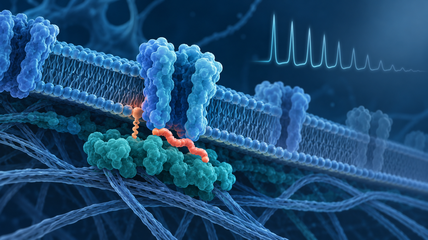
Conceptual illustration, not a measured molecular structure or study figure. The proposed chain is that disrupting the Nav1.8–Ank3 interaction reduces surface retention of Nav1.8 in sensitized nociceptors.
What are Nav1.8, ankyrin-3 and the SLiP?
Nav1.8 is a voltage-gated sodium channel expressed mainly in peripheral sensory neurons, including nociceptors. Sodium-channel activity helps these cells generate electrical signals. Nav1.8 is already an important pain-research target, but this study did not primarily try to plug the channel pore. It targeted a protein interaction that may help position the channel at the membrane.
Ankyrins are scaffold proteins that organize membrane proteins and connect them with the cell’s internal architecture. The authors found that the role of Ank3 was state-dependent: knocking down Ank3 reduced tetrodotoxin-resistant sodium conductance when cultured neurons had been pushed into a sensitized state, but not under basal conditions.
The experimental SLiP mimicked part of Nav1.8’s ankyrin-binding domain and carried a lipid group intended to help it associate with cell membranes. A scrambled-sequence lipidated peptide served as the main control. This construct is an experimental research tool, not an approved medicine or a clinically validated product.
Evidence layer 1: did the peptide disrupt biochemical binding?
Yes, the biochemical results support disruption of the Nav1.8–Ank3 complex—but that is a molecular endpoint. Co-immunoprecipitation experiments in mouse dorsal-root-ganglion and sciatic-nerve material found less Ank3 pulled down with Nav1.8 in the presence of the Nav1.8 peptide than with the scrambled control.
This matters because the peptide was designed around a specific interaction, and the result points in the intended direction. It does not show that every effect was caused only by that interaction, rule out other binding partners, or establish whole-organism selectivity.
Evidence layer 2: what happened in cultured sensory neurons?
The peptide’s electrical effects appeared mainly in sensitized or injury-associated neurons, not in ordinary basal conditions. That state dependence is central to the paper’s argument.
Across cultured mouse dorsal-root-ganglion experiments, the researchers reported that the Nav1.8 SLiP:
- reduced Nav1.8 surface fluorescence after cAMP-pathway activation, while a supplementary experiment did not show the same effect on Nav1.7;
- reduced total and tetrodotoxin-resistant sodium conductance in sensitized neurons;
- did not reduce conductance in Nav1.8-knockout neurons, supporting dependence on Nav1.8;
- reduced evoked firing and shifted measures such as rheobase toward a less excitable state; and
- reduced calcium responses in sensitized neuron-compartment experiments.
An inhibitor of clathrin-mediated endocytosis partly blunted the peptide’s effect in a supplementary experiment. That supports a model in which disrupting the scaffold allows Nav1.8 to be removed from the membrane. It is still a mechanistic interpretation, not a direct map of every trafficking step.
The selectivity evidence is encouraging but limited. Showing Nav1.8 dependence and no detected Nav1.7 surface reduction is not the same as a broad off-target screen, in-vivo toxicology program or complete assessment of other ion channels and proteins.
Evidence layer 3: what happened in mice?
The investigators used three injury or inflammation paradigms, all in young mice and all with local experimental exposure, not a human-ready systemic delivery strategy.
Neuroma model
After a surgically created sciatic-nerve neuroma was allowed to develop for eight weeks, the peptide was placed locally in a silicone cuff around the lesion. Compared with each animal’s pretreatment behavior and with scrambled-peptide controls, the study reported:
- fewer spontaneous hind-limb flicking bouts;
- fewer responses to mechanical stimulation of the neuroma, with separation emerging on day 4 and remaining significant through day 7; and
- fewer small-diameter sensory neurons responding during in-vivo calcium imaging when the neuroma was mechanically stimulated.
The neuroma behavioral cohorts were small. For example, the spontaneous-flick analysis included five scrambled-control and eight Nav1.8-SLiP mice; the mechanical-response analysis included four and five. Those are signals to replicate, not precise estimates of efficacy.
The paper also reported no reduction in locomotor distance at the peptide’s peak behavioral effect in five animals. A separate gabapentin experiment reduced locomotion. This was not a powered head-to-head trial of comparative efficacy or safety, so it should not be read as proof that the peptide is safer or better than gabapentin.
Spared-nerve-injury and inflammatory models
In the chronic spared-nerve-injury model, local hind-paw exposure was followed by higher mechanical withdrawal thresholds and less cold-evoked nocifensive behavior than with scrambled peptide. In the zymosan inflammatory model, the peptide group showed lower thermal and mechanical hypersensitivity after inflammation.
These experiments broaden the preclinical signal beyond one surgical model. They do not establish how long an effect would last beyond the reported windows, what repeated exposure would do, whether distant or systemic administration could reach the relevant neurons, or whether comparable exposure is feasible in people.
Was protective pain really spared?
The study supports a narrow version of that claim. In uninjured mice, the Nav1.8 SLiP did not change mechanical withdrawal thresholds measured 24 and 48 hours after local exposure. The supplementary experiment included four mice per group.
That is consistent with the state-dependent cell findings and with sparing this particular acute mechanical response over this short window. It does not prove that all protective nociception was preserved across thermal, chemical, visceral or longer-term challenges. “Did not significantly change” in a small experiment is also not proof of equivalence.
What the study does—and does not—show
Supported by the study:
- Nav1.8 and Ank3 can interact in mouse sensory-neuron material.
- The designed peptide disrupted that association in biochemical assays.
- In sensitized mouse neurons, the peptide reduced Nav1.8 membrane localization, Nav1.8-related conductance and hyperexcitability.
- Local exposure reduced selected pain-like behaviors in multiple mouse models.
- One short-term mechanical-nociception test in uninjured mice was unchanged.
Not established:
- pain relief or functional benefit in people;
- human safety, tolerability or an appropriate clinical exposure;
- long-term durability or effects of repeated exposure;
- comprehensive molecular selectivity or absence of off-target effects;
- a practical route, formulation, biodistribution profile or manufacturing strategy for clinical use; or
- superiority to an existing pain treatment.
This is why preclinical evidence should be read as a stage, not a synonym for “basically proven”. The mechanism can be coherent and the animal signal can be real while translation still fails.
The study’s main limitations
The authors explicitly identified several limitations, and the full paper plus supplement add more context.
The electrophysiology had technical constraints
Cultured sensory neurons extend processes that make voltage clamping difficult. The authors used reduced extracellular sodium and a pre-pulse protocol, but acknowledged residual space-clamp limitations and reported maximum conductance rather than full current-voltage curves. Adult neurons can make this problem worse because of their size and larger currents.
Some behavioral groups were small
Several neuroma comparisons used only four to nine animals per group. The larger supplementary injury-model cohorts improve context, but none substitute for independent replication, prespecified confirmatory studies or a development-grade safety program.
Sex was included but not resolved
Male and female mice were used, and the authors pooled them after observing no significant difference. Behavioral testing was performed in male cohorts and then female cohorts rather than interleaving sex. The paper acknowledges that a properly powered study could reveal sex-dependent responses missed here. “No observed difference” is not the same as evidence that sex does not matter.
Route and exposure were tightly experimental
The peptide was delivered locally near a neuroma or into the affected hind paw, and the cell studies used controlled laboratory exposure. These approaches are useful for testing mechanism. They do not answer pharmacokinetics, distribution, metabolism, immunogenicity, repeated-exposure safety, scalable formulation or whether enough intact peptide could reach human nociceptors through a practical clinical route.
The models are not neuropathic pain in people
Withdrawal thresholds, limb flicks and responses to cold or mechanical stimulation are animal behavioral endpoints. They are not human reports of spontaneous pain, sleep, function or quality of life. The mice were 5–7 weeks old and the experiments modeled selected injuries under controlled conditions, not the heterogeneity, comorbidities and medication histories seen in patients.
Human relevance remains hypothetical
The supplementary analysis showed Nav1.8 and ankyrin transcripts in human sensory-neuron datasets, and the paper notes that the relevant human and mouse proteins share substantial sequence identity. The authors were clear that the interaction’s importance across species still requires experimental validation. No human neurons, human tissue intervention, healthy volunteers or patients were treated with this peptide.
For a broader way to rank these claims, see Peptide Research Status Explained. A mechanism plus mouse behavior belongs on the evidence ladder; it does not jump over the human steps.
Funding, intellectual property and company ties
The paper’s declaration of interests reports that lead author Ryan Powell is chief scientific officer and co-founder of Mimetic Medicines. Clifford Woolf is on Mimetic Medicines’ scientific advisory board and has additional company relationships. Powell, Woolf and Xianhua Zhang reported a patent application related to the work filed through Boston Children’s Hospital. Other authors disclosed advisory relationships listed in the paper.
Boston Children’s Hospital also reported that it licensed the relevant intellectual property to Mimetic Medicines. These relationships do not make the findings false. They are relevant context when evaluating a translational claim tied to patented technology and a company developing it.
Bottom line
This study makes a strong preclinical case for a testable mechanism: in sensitized mouse nociceptors, Ank3 appears to help stabilize Nav1.8 at the membrane, and a lipidated peptide that disrupts the interaction can reduce channel availability, neuronal hyperexcitability and selected pain-like behaviors.
The evidence stops there. It does not yet tell us whether the approach can be delivered safely, selectively and durably to human sensory neurons—or whether doing so would improve neuropathic pain in people. That next gap is not paperwork. It is the experiment.
Sources
- Powell R, et al. Disruption of the Nav1.8/Ankyrin-3 interaction in nociceptors reduces pathological pain-like behaviors in mice. Cell Reports Medicine. Available online October 6, 2026.
- PubMed record and author conflict-of-interest statement.
- Boston Children’s Hospital. Researchers describe new approach to reducing neuropathic pain.
This article is educational and is not medical advice. It does not provide treatment, dosing, administration, purchasing, sourcing or self-experimentation guidance.