How Tissue-Damage Peptides May Block Lung Repair: What the New COPD Study Shows
A 2026 COPD study links elastin-derived peptides to impaired alveolar repair across human tissue, organoids, and mice—but the intervention evidence remains preclinical.
How Tissue-Damage Peptides May Block Lung Repair: What the New COPD Study Shows
Short answer: A 2026 study suggests that elastin-derived peptides may do more than mark tissue damage in chronic obstructive pulmonary disease (COPD). Across human lung samples, cigarette-smoke-exposed mice, and alveolar organoids, the researchers built a preclinical case that these fragments can interfere with the process by which repair-capable alveolar type 2 cells become thin, gas-exchanging alveolar type 1 cells. The work is mechanistically coherent, but it did not test a treatment in people.
The term peptide needs an immediate qualifier here. Elastin-derived peptides (EDPs) are fragments released when the structural protein elastin breaks down. They are endogenous damage products—not wellness peptides, supplements, or the research compounds commonly sold online.
Zhu and colleagues published the peer-reviewed study in MedComm on July 25, 2026. Its value is not that any one experiment “proved” a COPD therapy. Its value is that several different models pointed toward the same biological chain, while each model answered a different question.
What are elastin-derived peptides in COPD?
Elastin-derived peptides are bioactive fragments produced when elastin in the extracellular matrix is degraded. In the lung, elastin helps alveoli stretch and recoil. Cigarette-smoke-related inflammation and protease activity can damage this matrix, releasing fragments that may themselves affect cell signaling.
That distinction changes how the study should be read. The researchers were not adding a fashionable peptide to “optimize” lung function. They were asking whether the debris produced by damaged lung tissue could become part of the damage process.
This is biologically plausible because extracellular-matrix fragments are not always inert rubble. Some act as signaling molecules—sometimes called matrikines—that can influence inflammation, cell movement, or tissue remodeling. The new study investigated whether EDPs belong in that category during COPD-related alveolar injury.
Quick answer: EDPs are pieces of the body's own elastin released during matrix breakdown. “Peptide” describes their molecular size and composition; it does not make them equivalent to consumer peptide products.
What mechanism did the elastin-derived peptides COPD study propose?
The proposed pathway runs from matrix damage to a stalled repair program: cigarette-smoke-related elastin breakdown increases EDPs; EDP exposure activates TLR4/NF-κB-associated inflammatory signaling; DKK1 rises and β-catenin activity falls; alveolar type 2 (AT2) cells then have more difficulty differentiating into alveolar type 1 (AT1) cells.
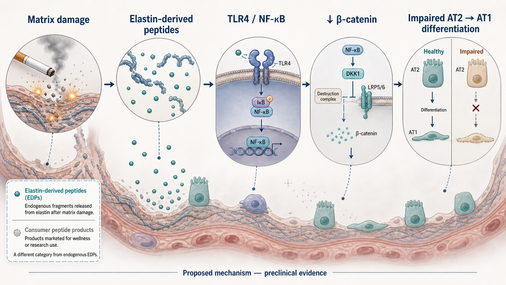
The study's proposed chain links elastin breakdown to inflammatory signaling and impaired alveolar-cell differentiation. This is a preclinical mechanism, not a demonstrated treatment pathway in patients.
AT2 and AT1 cells do different jobs. AT2 cells produce surfactant and can act as facultative progenitors after injury. AT1 cells form most of the thin surface across which gas exchange occurs. Repair therefore depends partly on AT2 cells moving through transitional states and becoming AT1 cells.
TLR4 is an innate-immune sensor, and NF-κB is a major inflammatory transcription pathway. β-catenin participates in canonical Wnt signaling, which helps regulate cell fate and regenerative programs. The paper reported increased DKK1—a Wnt-pathway inhibitor—alongside reduced β-catenin activity. That gives the model a plausible bridge between an inflammatory signal and a weaker differentiation program.
The important word is proposed. A pathway diagram can make biology look like plumbing: one pipe enters, one valve turns, and one outcome emerges. Living tissue is messier. COPD involves smoke exposure, immune cells, proteases, oxidative stress, infection risk, vascular changes, and many interacting repair pathways. The study supports EDPs as one contributor, not the sole explanation.
What did the human COPD tissue show?
The human evidence established disease relevance and association, not treatment efficacy or complete causation. The researchers analyzed lung tissue from eight people with moderate-to-severe COPD and eight age-matched controls without airflow obstruction. COPD samples showed emphysematous changes, disrupted elastic fibers, altered epithelial-cell states, and higher relative EDP levels in serum and bronchoalveolar lavage fluid.
Single-cell RNA sequencing and pseudotime analyses were consistent with impaired AT2-to-AT1 differentiation. The study also reported fewer AT1 cells and changes in transitional epithelial populations in COPD samples.
These observations matter because they show that the proposed biology is present in human disease tissue. But tissue association cannot establish that EDPs caused the disease pattern. The serum and lavage samples used for EDP measurement came from independent, unpaired clinical cohorts rather than the same individuals who provided surgical tissue, so the human findings are best read as group-level associations.
Evidence boundary: Human tissue made the mechanism relevant to COPD. It did not show that blocking EDPs repairs human lungs, improves breathing, reduces exacerbations, or changes any patient outcome.
What did the cigarette-smoke mouse model add?
The mouse experiments showed that the same broad pattern appeared in a controlled whole-animal model. After four months of cigarette-smoke exposure, mice displayed emphysema-like structural changes, impaired pulmonary-function measures, disrupted elastin, higher relative EDP levels, and evidence of impaired AT2-to-AT1 differentiation.
This layer is stronger than a cell dish alone because it preserves interactions among the epithelium, immune system, circulation, and extracellular matrix. It also allowed the researchers to examine an intervention in a living organism.
The tradeoff is translation. A smoke-exposed mouse is a model of selected COPD-like features, not a person living with heterogeneous, long-standing COPD. Mouse responses can identify a mechanism worth pursuing without predicting whether a human intervention will be effective or safe.
What did the human and mouse alveolar organoids test?
Organoids supplied the study's clearest controlled functional test. These three-dimensional cultures model parts of alveolar growth and differentiation while allowing researchers to change one exposure at a time.
In both mouse and human alveolar organoids, cigarette-smoke extract and added EDPs suppressed organoid growth and impaired AT2-to-AT1 differentiation. Finding a similar response in organoids derived from two species reduces the chance that the result is a quirk of one model.
It still does not recreate a complete lung. Organoids lack normal breathing mechanics, circulation, full immune interactions, and the decades-long disease history that can shape COPD. Their strength is controlled mechanism testing, not forecasting clinical benefit.
Quick answer: The organoids showed that EDP exposure could alter alveolar-cell behavior under controlled conditions. They did not show that EDPs are the only cause of failed repair in COPD.
How did pathway perturbation strengthen the case?
Interfering with the proposed pathway made the study more informative than a correlation-only paper. EDP exposure was associated with greater TLR4/NF-κB activity and lower β-catenin activity. Pharmacologically inhibiting TLR4 partially restored alveolar epithelial differentiation in the organoid experiments.
The word partially is important. Partial restoration is consistent with TLR4 signaling contributing to the effect, while also suggesting that other pathways or damage processes remain involved. The authors noted that additional inflammatory signals may matter, and they called for further genetic and mechanistic work to establish causal signaling relationships.
This kind of perturbation asks a better question than “were two markers present together?” If changing a proposed node also changes the downstream phenotype, the pathway becomes more credible. It is still possible for a drug inhibitor to have off-target effects, and pharmacological perturbation is not the same as definitive genetic proof.
What happened with the experimental EDP-neutralizing agent?
The agent TB-B002D, abbreviated TB in the paper, produced a partial rescue in organoids and reduced several emphysema-like findings in mice. TB is a peptide-based experimental agent designed to neutralize excessive EDPs. In organoids exposed to cigarette-smoke extract or EDPs, it partially restored growth and AT2-to-AT1 differentiation. In smoke-exposed mice, it was associated with better histological and selected lung-function measures than smoke exposure alone.
This is intervention evidence within preclinical models. It supports the idea that EDPs may be functionally involved rather than merely present after damage.
It is not evidence that TB benefits people with COPD. The agent is not an approved therapy, and the paper did not report a human treatment trial. Its molecular specificity in COPD, long-term safety, durability, delivery, and translational feasibility remain unresolved. The company that provided TB was acknowledged in the paper; the authors declared no conflicts of interest.
Bottom line on the rescue: Reversing part of a model phenotype is stronger evidence than observing a marker. It remains several steps away from proving clinical benefit.
Why is this evidence package stronger than a single cell assay?
The study gains strength from convergence: different methods, each with different weaknesses, pointed toward the same model. No single layer carries the conclusion by itself.
| Evidence layer | What it adds | What it cannot establish | |---|---|---| | Human COPD tissue and fluids | Disease relevance; EDP elevation and repair defects occur in human COPD groups | Individual-level causation or treatment benefit | | Smoke-exposed mice | A whole-organism model with matrix, immune, epithelial, and functional measurements | Human efficacy or safety | | Human and mouse alveolar organoids | Controlled tests of EDP effects on growth and differentiation | The full complexity of a living lung | | TLR4 inhibition and pathway measurements | Evidence that the proposed signaling chain is functionally connected | Complete pathway specificity or sole causation | | TB rescue in organoids and mice | Intervention support for EDP involvement | Clinical benefit, approval, or readiness for patient use |
This is what a mechanistically coherent translational package looks like: human observations establish relevance; animal work supplies an intact biological system; organoids isolate function; pathway perturbation tests the mechanism; and rescue experiments ask whether changing the target changes the phenotype.
That package is more persuasive than one cell assay because its pieces fail in different ways. Agreement across them makes a coincidence or model-specific artifact less likely. It does not remove the preclinical boundary. For a broader guide to that boundary, see What Preclinical Actually Means in Peptide Research and Peptide Research Status Explained.
What are the study's biggest limitations?
The central limitation is simple: nobody with COPD received an EDP-neutralizing treatment in this study. Everything about rescue or intervention came from organoids or mice.
Other limitations include:
- The human surgical comparison involved only eight COPD samples and eight controls.
- Human EDP measurements and surgical tissue analyses came from independent, unpaired cohorts.
- Tissue associations cannot prove that EDPs caused the observed differentiation defect.
- The downstream signaling mechanism was only partly resolved and needs genetic confirmation.
- The mouse treatment experiment was short-term and used one specific experimental model.
- TB's specificity, long-term safety, durability, delivery, and feasibility in humans are unknown.
- COPD is biologically heterogeneous, so one matrix-derived pathway is unlikely to explain every patient or disease stage.
There is also a language trap in the paper's title: “reverses alveolar epithelial dysfunction” describes experimental findings in model systems. It should not be shortened into “reverses COPD” or “repairs human lungs.” Those would be different claims requiring clinical evidence.
What would need to happen next?
The next steps are replication, sharper mechanism testing, and staged safety work—not clinical conclusions by press release. Independent groups would need to reproduce the findings, clarify which EDP species matter, test target specificity, examine longer exposures, and determine whether the biology holds across more diverse human samples.
If an EDP-targeted agent eventually advances toward human research, investigators would first need to characterize manufacturing, pharmacology, delivery, toxicology, and exposure behavior before asking whether it improves meaningful COPD outcomes. A true efficacy trial would need outcomes that matter to patients, not only pathway markers or tissue images.
The bottom line
Zhu et al. provide a credible preclinical case that elastin-derived peptides may help connect tissue destruction to failed alveolar repair in COPD. The strongest feature is the alignment of human observations, mouse biology, cross-species organoid experiments, pathway perturbation, and partial rescue.
The study does not establish EDPs as the sole driver of COPD, and it does not show that neutralizing them helps patients. TB-B002D remains experimental and unapproved. The responsible conclusion is neither “just a cell study” nor “a new COPD treatment.” It is a well-layered translational study that makes a mechanism worth testing more rigorously.
This article is for general education and evidence literacy. It is not medical advice and does not diagnose COPD, recommend a treatment, or advise changing medical care.
Primary source
Zhu H, Zhao Y, Qin Y, et al. Targeting Elastin-Derived Peptides Reverses Alveolar Epithelial Dysfunction in Chronic Obstructive Pulmonary Disease. MedComm. Published online July 25, 2026. PubMed PMID: 42502713; PMCID: PMC13401145.